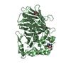
[English] 日本語
Yorodumi
- PDB-3fie: Crystal structure of Clostridium botulinum neurotoxin serotype F ... -

+
Open data


ID or keywords:

Loading...

-
Basic information

Entry
Database: PDB / ID: 3fie
TitleCrystal structure of Clostridium botulinum neurotoxin serotype F catalytic domain with an inhibitor (inh1)
Components
  • BOTULINUM NEUROTOXIN TYPE F
  • fragment of Vesicle-associated membrane protein 2
KeywordsHYDROLASE / TOXIN/PROTEIN TRANSPORT / Clostridium botulinum / BoNT F / VAMP / inhibitor / complex structure / Acetylation / Cell junction / TOXIN-PROTEIN TRANSPORT COMPLEX
Function / homology
Function and homology information


Toxicity of tetanus toxin (tetX) / clathrin-sculpted glutamate transport vesicle membrane / Toxicity of botulinum toxin type F (botF) / Toxicity of botulinum toxin type G (botG) / Toxicity of botulinum toxin type D (botD) / regulation of delayed rectifier potassium channel activity / trans-Golgi Network Vesicle Budding / Acetylcholine Neurotransmitter Release Cycle / synaptobrevin 2-SNAP-25-syntaxin-1a-complexin II complex / synaptobrevin 2-SNAP-25-syntaxin-1a complex ...Toxicity of tetanus toxin (tetX) / clathrin-sculpted glutamate transport vesicle membrane / Toxicity of botulinum toxin type F (botF) / Toxicity of botulinum toxin type G (botG) / Toxicity of botulinum toxin type D (botD) / regulation of delayed rectifier potassium channel activity / trans-Golgi Network Vesicle Budding / Acetylcholine Neurotransmitter Release Cycle / synaptobrevin 2-SNAP-25-syntaxin-1a-complexin II complex / synaptobrevin 2-SNAP-25-syntaxin-1a complex / synaptobrevin 2-SNAP-25-syntaxin-1a-complexin I complex / clathrin-sculpted monoamine transport vesicle membrane / Toxicity of botulinum toxin type B (botB) / clathrin-sculpted gamma-aminobutyric acid transport vesicle membrane / zymogen granule membrane / Serotonin Neurotransmitter Release Cycle / GABA synthesis, release, reuptake and degradation / Dopamine Neurotransmitter Release Cycle / eosinophil degranulation / Norepinephrine Neurotransmitter Release Cycle / vesicle fusion / SNARE complex / Golgi to plasma membrane protein transport / SNAP receptor activity / regulation of vesicle-mediated transport / Glutamate Neurotransmitter Release Cycle / bontoxilysin / calcium-ion regulated exocytosis / positive regulation of intracellular protein transport / host cell presynaptic membrane / regulation of exocytosis / host cell cytoplasmic vesicle / neuron projection terminus / Sensory processing of sound by inner hair cells of the cochlea / syntaxin-1 binding / SNARE complex assembly / syntaxin binding / host cell cytosol / clathrin-coated vesicle / Lysosome Vesicle Biogenesis / Insertion of tail-anchored proteins into the endoplasmic reticulum membrane / Other interleukin signaling / Golgi Associated Vesicle Biogenesis / Insulin processing / exocytosis / synaptic vesicle exocytosis / synaptic vesicle endocytosis / protein transmembrane transporter activity / response to glucose / vesicle-mediated transport / SNARE binding / secretory granule membrane / secretory granule / long-term synaptic potentiation / Regulation of insulin secretion / Translocation of SLC2A4 (GLUT4) to the plasma membrane / phospholipid binding / clathrin-coated endocytic vesicle membrane / trans-Golgi network / synaptic vesicle membrane / metalloendopeptidase activity / cellular response to insulin stimulus / calcium-dependent protein binding / synaptic vesicle / protein transport / Cargo recognition for clathrin-mediated endocytosis / Clathrin-mediated endocytosis / toxin activity / cytoplasmic vesicle / protein-containing complex assembly / vesicle / membrane fusion / calmodulin binding / neuron projection / intracellular membrane-bounded organelle / synapse / lipid binding / host cell plasma membrane / perinuclear region of cytoplasm / proteolysis / zinc ion binding / extracellular region / membrane / plasma membrane / cytosol
Similarity search - Function
Synaptobrevin/Vesicle-associated membrane protein / Synaptobrevin signature. / Synaptobrevin-like / Synaptobrevin / v-SNARE, coiled-coil homology domain / v-SNARE coiled-coil homology domain profile. / Zincin-like / Metalloproteases ("zincins"), catalytic domain like / Clostridium neurotoxin, translocation / Clostridium neurotoxin, Translocation domain ...Synaptobrevin/Vesicle-associated membrane protein / Synaptobrevin signature. / Synaptobrevin-like / Synaptobrevin / v-SNARE, coiled-coil homology domain / v-SNARE coiled-coil homology domain profile. / Zincin-like / Metalloproteases ("zincins"), catalytic domain like / Clostridium neurotoxin, translocation / Clostridium neurotoxin, Translocation domain / Clostridium neurotoxin, translocation domain / Clostridial neurotoxin zinc protease / Botulinum/Tetanus toxin, catalytic chain / Clostridium neurotoxin, receptor binding N-terminal / Clostridium neurotoxin, receptor-binding C-terminal / Clostridium neurotoxin, C-terminal receptor binding / Clostridium neurotoxin, N-terminal receptor binding / Kunitz inhibitor STI-like superfamily / Neutral zinc metallopeptidases, zinc-binding region signature. / Concanavalin A-like lectin/glucanase domain superfamily / Alpha-Beta Complex / Alpha Beta
Similarity search - Domain/homology
Botulinum neurotoxin type F / Vesicle-associated membrane protein 2 / Neurotoxin type F
Similarity search - Component
Biological speciesClostridium botulinum (bacteria)
MethodX-RAY DIFFRACTION / SYNCHROTRON / MOLECULAR REPLACEMENT / Resolution: 2.1 Å
AuthorsAgarwal, R. / Swaminathan, S.
CitationJournal: Nat.Struct.Mol.Biol. / Year: 2009
Title: Mode of VAMP substrate recognition and inhibition of Clostridium botulinum neurotoxin F.
Authors: Agarwal, R. / Schmidt, J.J. / Stafford, R.G. / Swaminathan, S.
History
DepositionDec 11, 2008Deposition site: RCSB / Processing site: RCSB
Revision 1.0Jun 23, 2009Provider: repository / Type: Initial release
Revision 1.1Jul 13, 2011Group: Version format compliance
Revision 1.2Sep 6, 2023Group: Data collection / Database references ...Data collection / Database references / Derived calculations / Refinement description
Category: chem_comp_atom / chem_comp_bond ...chem_comp_atom / chem_comp_bond / database_2 / pdbx_initial_refinement_model / struct_conn / struct_ref_seq_dif / struct_site
Item: _database_2.pdbx_DOI / _database_2.pdbx_database_accession ..._database_2.pdbx_DOI / _database_2.pdbx_database_accession / _struct_conn.conn_type_id / _struct_conn.id / _struct_conn.pdbx_dist_value / _struct_conn.pdbx_leaving_atom_flag / _struct_conn.ptnr1_auth_asym_id / _struct_conn.ptnr1_auth_comp_id / _struct_conn.ptnr1_auth_seq_id / _struct_conn.ptnr1_label_asym_id / _struct_conn.ptnr1_label_atom_id / _struct_conn.ptnr1_label_comp_id / _struct_conn.ptnr1_label_seq_id / _struct_conn.ptnr2_auth_asym_id / _struct_conn.ptnr2_auth_comp_id / _struct_conn.ptnr2_auth_seq_id / _struct_conn.ptnr2_label_asym_id / _struct_conn.ptnr2_label_atom_id / _struct_conn.ptnr2_label_comp_id / _struct_conn.ptnr2_label_seq_id / _struct_ref_seq_dif.details / _struct_site.pdbx_auth_asym_id / _struct_site.pdbx_auth_comp_id / _struct_site.pdbx_auth_seq_id

-
Structure visualization

Structure viewerMolecule:
MolmilJmol/JSmol

Downloads & links

-
Assembly

Deposited unit
A: BOTULINUM NEUROTOXIN TYPE F
B: BOTULINUM NEUROTOXIN TYPE F
C: fragment of Vesicle-associated membrane protein 2
D: fragment of Vesicle-associated membrane protein 2
hetero molecules


Theoretical massNumber of molelcules
Total (without water)106,1846
Polymers106,0534
Non-polymers1312
Water6,287349
1
A: BOTULINUM NEUROTOXIN TYPE F
C: fragment of Vesicle-associated membrane protein 2
hetero molecules


Theoretical massNumber of molelcules
Total (without water)53,0923
Polymers53,0262
Non-polymers651
Water362
TypeNameSymmetry operationNumber
identity operation1_555x,y,z1
Buried area3980 Å2
ΔGint-53 kcal/mol
Surface area18310 Å2
MethodPISA
2
B: BOTULINUM NEUROTOXIN TYPE F
D: fragment of Vesicle-associated membrane protein 2
hetero molecules


Theoretical massNumber of molelcules
Total (without water)53,0923
Polymers53,0262
Non-polymers651
Water362
TypeNameSymmetry operationNumber
identity operation1_555x,y,z1
Buried area3800 Å2
ΔGint-56 kcal/mol
Surface area18100 Å2
MethodPISA
Unit cell
Length a, b, c (Å)55.637, 61.389, 70.027
Angle α, β, γ (deg.)94.24, 89.92, 113.27
Int Tables number1
Space group name H-MP1

-
Components

#1: Protein BOTULINUM NEUROTOXIN TYPE F / E.C.3.4.24.69 / BoNT/F (Neurotoxin type F)


Mass: 48745.621 Da / Num. of mol.: 2 / Fragment: residues 1-419, catalytic domain
Source method: isolated from a genetically manipulated source
Source: (gene. exp.) Clostridium botulinum (bacteria) / Gene: bonT/F / Plasmid: pET28b / Production host: Escherichia coli (E. coli) / Strain (production host): BL21(DE3)RIPL
References: UniProt: Q57236, UniProt: A7GBG3*PLUS, bontoxilysin
#2: Protein/peptide fragment of Vesicle-associated membrane protein 2 / VAMP-2 / Synaptobrevin-2


Mass: 4280.842 Da / Num. of mol.: 2 / Fragment: residues 22-58 / Mutation: Q58DCY / Source method: obtained synthetically / Details: Sequence occurs in Homo sapiens / References: UniProt: P63027
#3: Chemical ChemComp-ZN / ZINC ION


Mass: 65.409 Da / Num. of mol.: 2 / Source method: obtained synthetically / Formula: Zn
#4: Water ChemComp-HOH / water / Water


Mass: 18.015 Da / Num. of mol.: 349 / Source method: isolated from a natural source / Formula: H2O

-
Experimental details

-
Experiment

ExperimentMethod: X-RAY DIFFRACTION / Number of used crystals: 1

-
Sample preparation

CrystalDensity Matthews: 2.07 Å3/Da / Density % sol: 40.44 %
Crystal growTemperature: 298 K / Method: vapor diffusion, sitting drop / pH: 5.5
Details: 0.1M Bis-tris, 25% PEG 3350, pH 5.5, VAPOR DIFFUSION, SITTING DROP, temperature 298K

-
Data collection

DiffractionMean temperature: 100 K
Diffraction sourceSource: SYNCHROTRON / Site: NSLS / Beamline: X12C / Wavelength: 1.08 Å
DetectorType: ADSC QUANTUM 210 / Detector: CCD / Date: Jun 11, 2008 / Details: mirrors
RadiationMonochromator: si(iii) / Protocol: SINGLE WAVELENGTH / Monochromatic (M) / Laue (L): M / Scattering type: x-ray
Radiation wavelengthWavelength: 1.08 Å / Relative weight: 1
ReflectionResolution: 2.1→40.65 Å / Num. all: 48376 / Num. obs: 48376 / % possible obs: 96.5 % / Observed criterion σ(F): 0 / Observed criterion σ(I): 0 / Redundancy: 3.7 % / Biso Wilson estimate: 12.3 Å2 / Rmerge(I) obs: 0.06 / Net I/σ(I): 14.4
Reflection shellResolution: 2.1→2.18 Å / Redundancy: 3.2 % / Rmerge(I) obs: 0.19 / Num. unique all: 4368 / % possible all: 87.2

-
Processing

Software
NameVersionClassification
CBASSdata collection
MOLREPphasing
Cootmodel building
CNS1.1refinement
HKL-2000data reduction
HKL-2000data scaling
SCALEPACKdata scaling
RefinementMethod to determine structure: MOLECULAR REPLACEMENT
Starting model: 2A8A
Resolution: 2.1→40.65 Å / Rfactor Rfree error: 0.007 / Data cutoff high absF: 123485.81 / Data cutoff low absF: 0 / Isotropic thermal model: RESTRAINED / Cross valid method: THROUGHOUT / σ(F): 0 / Stereochemistry target values: Engh & Huber
RfactorNum. reflection% reflectionSelection details
Rfree0.279 1420 3 %RANDOM
Rwork0.228 ---
all0.2281 48376 --
obs0.2281 47160 95.2 %-
Solvent computationSolvent model: FLAT MODEL / Bsol: 42.6656 Å2 / ksol: 0.35665 e/Å3
Displacement parametersBiso mean: 22.6 Å2
Baniso -1Baniso -2Baniso -3
1--2.3 Å23.16 Å2-0.55 Å2
2--3.31 Å21.51 Å2
3----1 Å2
Refine analyze
FreeObs
Luzzati coordinate error0.35 Å0.27 Å
Luzzati d res low-5 Å
Luzzati sigma a0.35 Å0.21 Å
Refinement stepCycle: LAST / Resolution: 2.1→40.65 Å
ProteinNucleic acidLigandSolventTotal
Num. atoms7039 0 2 349 7390
Refine LS restraints
Refine-IDTypeDev ideal
X-RAY DIFFRACTIONc_bond_d0.007
X-RAY DIFFRACTIONc_angle_deg1.4
X-RAY DIFFRACTIONc_dihedral_angle_d23.1
X-RAY DIFFRACTIONc_improper_angle_d0.85
LS refinement shellResolution: 2.1→2.23 Å / Rfactor Rfree error: 0.023 / Total num. of bins used: 6
RfactorNum. reflection% reflection
Rfree0.346 227 3 %
Rwork0.271 7226 -
obs-48376 90.1 %
Xplor file
Refine-IDSerial noParam fileTopol file
X-RAY DIFFRACTION1protein_rep.paramprotein.top
X-RAY DIFFRACTION2ion.paramdcys.top-org
X-RAY DIFFRACTION3water_rep.param
X-RAY DIFFRACTION4dcys.param-org

+
About Yorodumi

-
News

-
Feb 9, 2022. New format data for meta-information of EMDB entries

New format data for meta-information of EMDB entries

  • Version 3 of the EMDB header file is now the official format.
  • The previous official version 1.9 will be removed from the archive.

Related info.:EMDB header

External links:wwPDB to switch to version 3 of the EMDB data model

-
Aug 12, 2020. Covid-19 info

Covid-19 info

URL: https://pdbj.org/emnavi/covid19.php

New page: Covid-19 featured information page in EM Navigator.

Related info.:Covid-19 info / Mar 5, 2020. Novel coronavirus structure data

+
Mar 5, 2020. Novel coronavirus structure data

Novel coronavirus structure data

Related info.:Yorodumi Speices / Aug 12, 2020. Covid-19 info

External links:COVID-19 featured content - PDBj / Molecule of the Month (242):Coronavirus Proteases

+
Jan 31, 2019. EMDB accession codes are about to change! (news from PDBe EMDB page)

EMDB accession codes are about to change! (news from PDBe EMDB page)

  • The allocation of 4 digits for EMDB accession codes will soon come to an end. Whilst these codes will remain in use, new EMDB accession codes will include an additional digit and will expand incrementally as the available range of codes is exhausted. The current 4-digit format prefixed with “EMD-” (i.e. EMD-XXXX) will advance to a 5-digit format (i.e. EMD-XXXXX), and so on. It is currently estimated that the 4-digit codes will be depleted around Spring 2019, at which point the 5-digit format will come into force.
  • The EM Navigator/Yorodumi systems omit the EMD- prefix.

Related info.:Q: What is EMD? / ID/Accession-code notation in Yorodumi/EM Navigator

External links:EMDB Accession Codes are Changing Soon! / Contact to PDBj

+
Jul 12, 2017. Major update of PDB

Major update of PDB

  • wwPDB released updated PDB data conforming to the new PDBx/mmCIF dictionary.
  • This is a major update changing the version number from 4 to 5, and with Remediation, in which all the entries are updated.
  • In this update, many items about electron microscopy experimental information are reorganized (e.g. em_software).
  • Now, EM Navigator and Yorodumi are based on the updated data.

External links:wwPDB Remediation / Enriched Model Files Conforming to OneDep Data Standards Now Available in the PDB FTP Archive

-
Yorodumi

Thousand views of thousand structures

  • Yorodumi is a browser for structure data from EMDB, PDB, SASBDB, etc.
  • This page is also the successor to EM Navigator detail page, and also detail information page/front-end page for Omokage search.
  • The word "yorodu" (or yorozu) is an old Japanese word meaning "ten thousand". "mi" (miru) is to see.

Related info.:EMDB / PDB / SASBDB / Comparison of 3 databanks / Yorodumi Search / Aug 31, 2016. New EM Navigator & Yorodumi / Yorodumi Papers / Jmol/JSmol / Function and homology information / Changes in new EM Navigator and Yorodumi

Read more