 Movie
Movie Controller

[English] 日本語
 Yorodumi
- PDB-3fe3: Crystal structure of the kinase MARK3/Par-1: T211A-S215A double mutant -
+ Open data

- Basic information
| Entry | Database: PDB / ID: 3fe3 | ||||||
|---|---|---|---|---|---|---|---|
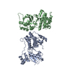
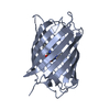
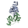
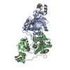
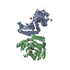
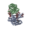
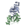
| Title | Crystal structure of the kinase MARK3/Par-1: T211A-S215A double mutant | ||||||
 Components Components | MAP/microtubule affinity-regulating kinase 3 | ||||||
 Keywords Keywords |  TRANSFERASE / SERINE/THREONINE PROTEIN KINASE / MARK / PAR-1 / KIN1 / UBA DOMAIN / C-TAK1 / P78 / MARK3 / ATP-binding / Kinase / Nucleotide-binding / Phosphoprotein / Serine/threonine-protein kinase TRANSFERASE / SERINE/THREONINE PROTEIN KINASE / MARK / PAR-1 / KIN1 / UBA DOMAIN / C-TAK1 / P78 / MARK3 / ATP-binding / Kinase / Nucleotide-binding / Phosphoprotein / Serine/threonine-protein kinase | ||||||
| Function / homology |  Function and homology information Function and homology informationpeptidyl-serine autophosphorylation / negative regulation of protein localization to nucleus / tau-protein kinase activity / negative regulation of hippo signaling / RAF activation / Signaling by high-kinase activity BRAF mutants / MAP2K and MAPK activation / tau protein binding / Negative regulation of MAPK pathway / Signaling by RAF1 mutants ...peptidyl-serine autophosphorylation / negative regulation of protein localization to nucleus / tau-protein kinase activity / negative regulation of hippo signaling / RAF activation / Signaling by high-kinase activity BRAF mutants / MAP2K and MAPK activation / tau protein binding / Negative regulation of MAPK pathway / Signaling by RAF1 mutants / Signaling by moderate kinase activity BRAF mutants / Paradoxical activation of RAF signaling by kinase inactive BRAF / Signaling downstream of RAS mutants / Signaling by BRAF and RAF1 fusions / positive regulation of protein binding / peptidyl-serine phosphorylation / non-specific serine/threonine protein kinase / intracellular signal transduction / protein phosphorylation / protein serine kinase activity / protein serine/threonine kinase activity / dendrite / extracellular exosome / ATP binding / plasma membrane / cytosol / cytoplasmSimilarity search - Function | ||||||
| Biological species |  Homo sapiens (human) Homo sapiens (human) | ||||||
| Method | X-RAY DIFFRACTION / SYNCHROTRON / MOLECULAR REPLACEMENT / molecular replacement / Resolution: 1.9 Å | ||||||
 Authors Authors | Nugoor, C. / Marx, A. / Panneerselvam, S. / Mandelkow, E.-M. / Mandelkow, E. | ||||||
 Citation Citation | Journal: TO BE PUBLISHED Title: Crystal structure of the kinase MARK3/Par-1: T211A-S215A double mutant Authors: Nugoor, C. / Marx, A. / Panneerselvam, S. / Mandelkow, E.-M. / Mandelkow, E. #1: Journal: Structure / Year: 2006Title: Structure of the catalytic and ubiquitin-associated domains of the protein kinase MARK/Par-1 Authors: Panneerselvam, S. / Marx, A. / Mandelkow, E.-M. / Mandelkow, E. #2: Journal: J.Biol.Chem. / Year: 2006Title: Structural variations in the catalytic and ubiquitin-associated domains of microtubule-associated protein/microtubule affinity regulating kinase (MARK) 1 and MARK2 Authors: Marx, A. / Nugoor, C. / Muller, J. / Panneerselvam, S. / Timm, T. / Bilang, M. / Mylonas, E. / Svergun, D.I. / Mandelkow, E.-M. / Mandelkow, E. | ||||||
| History |
|
- Structure visualization
| Structure viewer | Molecule: MolmilJmol/JSmol |
|---|
- Downloads & links
-Download
| PDBx/mmCIF format | 3fe3.cif.gz | 150.1 KB | Display | PDBx/mmCIF format |
|---|---|---|---|---|
| PDB format | pdb3fe3.ent.gz | 116.6 KB | Display | PDB format |
| PDBx/mmJSON format | 3fe3.json.gz | Tree view | PDBx/mmJSON format | |
| Others |  Other downloads Other downloads |
-Validation report
| Arichive directory | https://data.pdbj.org/pub/pdb/validation_reports/fe/3fe3ftp://data.pdbj.org/pub/pdb/validation_reports/fe/3fe3 | HTTPS FTP |
|---|
-Related structure data
| Related structure data | 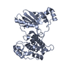 2hakS S: Starting model for refinement |
|---|---|
| Similar structure data |


-Links
 PDBj
PDBj- Assembly
| Deposited unit | 
| ||||||||
|---|---|---|---|---|---|---|---|---|---|
| 1 |
| ||||||||
| Unit cell |
| ||||||||
| Details | AUTHOR DETERMINED BIOLOGICAL UNIT: UNKNOWN |
-Components
| #1: Protein | Mass: 37619.582 Da / Num. of mol.: 2 Fragment: CATALYTIC AND UBIQUITIN-ASSOCIATED DOMAINS, UNP residues 41-367 Mutation: T211A, S215A Source method: isolated from a genetically manipulated source Source: (gene. exp.) Homo sapiens (human) / Gene: MARK3 / Plasmid: PET16b / Production host:  Escherichia coli (E. coli) / Strain (production host): BL21AI Escherichia coli (E. coli) / Strain (production host): BL21AIReferences: UniProt: P27448, non-specific serine/threonine protein kinase#2: Water | ChemComp-HOH / | Water Mass: 18.015 Da / Num. of mol.: 398 / Source method: isolated from a natural source / Formula: H2O Mass: 18.015 Da / Num. of mol.: 398 / Source method: isolated from a natural source / Formula: H2OSequence details | THE SEQUENCE IS BASED ON THE ISOFORM 3 OF DATABASE MARK3_HUMAN. RESIDUES UNP 162-184 ARE MISSING IN ISOFORM 3. | |
|---|
-Experimental details
-Experiment
| Experiment | Method: X-RAY DIFFRACTION / Number of used crystals: 1 |
|---|
- Sample preparation
| Crystal | Density Matthews: 3.26 Å3/Da / Density % sol: 62.25 % |
|---|---|
| Crystal grow | Temperature: 277 K / Method: vapor diffusion, hanging drop / pH: 7.5 Details: 100mM Hepes, 200mM calcium chloride, 15-18% PEG 3350, 5mM DTT , pH 7.5, VAPOR DIFFUSION, HANGING DROP, temperature 277K |
-Data collection
| Diffraction | Mean temperature: 100 K |
|---|---|
| Diffraction source | Source: SYNCHROTRON / Site: BESSY  / Beamline: 14.2 / Wavelength: 0.91841 Å / Beamline: 14.2 / Wavelength: 0.91841 Å |
| Detector | Type: MX-225 / Detector: CCD / Date: Jul 13, 2008 |
| Radiation | Protocol: SINGLE WAVELENGTH / Monochromatic (M) / Laue (L): M / Scattering type: x-ray |
| Radiation wavelength | Wavelength: 0.91841 Å / Relative weight: 1 |
| Reflection | Resolution: 1.9→62.3 Å / Num. obs: 75983 / % possible obs: 99.93 % / Redundancy: 4.95 % / Rmerge(I) obs: 0.037 / Net I/σ(I): 20.89 |
| Reflection shell | Resolution: 1.9→1.95 Å / Redundancy: 3.71 % / Rmerge(I) obs: 0.405 / Mean I/σ(I) obs: 3.51 / Num. unique all: 5590 / % possible all: 99.6 |
-Phasing
| Phasing | Method: molecular replacement |
|---|
- Processing
| Software |
| |||||||||||||||||||||||||||||||||||||||||||||||||||||||||||||||||||||||||||||||||||||||||||||||||||||||||||||||||||||||||||||||||||||||||||||||||||||||||||||||||||||||||||||||
|---|---|---|---|---|---|---|---|---|---|---|---|---|---|---|---|---|---|---|---|---|---|---|---|---|---|---|---|---|---|---|---|---|---|---|---|---|---|---|---|---|---|---|---|---|---|---|---|---|---|---|---|---|---|---|---|---|---|---|---|---|---|---|---|---|---|---|---|---|---|---|---|---|---|---|---|---|---|---|---|---|---|---|---|---|---|---|---|---|---|---|---|---|---|---|---|---|---|---|---|---|---|---|---|---|---|---|---|---|---|---|---|---|---|---|---|---|---|---|---|---|---|---|---|---|---|---|---|---|---|---|---|---|---|---|---|---|---|---|---|---|---|---|---|---|---|---|---|---|---|---|---|---|---|---|---|---|---|---|---|---|---|---|---|---|---|---|---|---|---|---|---|---|---|---|---|---|
| Refinement | Method to determine structure: MOLECULAR REPLACEMENT Starting model: PDB ENTRY 2HAK-E (52-304) Resolution: 1.9→51.85 Å / Cor.coef. Fo:Fc: 0.96 / Cor.coef. Fo:Fc free: 0.943 / WRfactor Rfree: 0.245 / WRfactor Rwork: 0.216 / Occupancy max: 1 / Occupancy min: 0.5 / SU B: 5.944 / SU ML: 0.093 / TLS residual ADP flag: LIKELY RESIDUAL / Isotropic thermal model: ISOTROPIC, TLS / Cross valid method: THROUGHOUT / σ(F): 0 / ESU R: 0.128 / ESU R Free: 0.125 / Stereochemistry target values: MAXIMUM LIKELIHOOD Details: HYDROGENS HAVE BEEN ADDED IN THE RIDING POSITIONS U VALUES: RESIDUAL ONLY
| |||||||||||||||||||||||||||||||||||||||||||||||||||||||||||||||||||||||||||||||||||||||||||||||||||||||||||||||||||||||||||||||||||||||||||||||||||||||||||||||||||||||||||||||
| Solvent computation | Ion probe radii: 0.8 Å / Shrinkage radii: 0.8 Å / VDW probe radii: 1.2 Å / Solvent model: MASK | |||||||||||||||||||||||||||||||||||||||||||||||||||||||||||||||||||||||||||||||||||||||||||||||||||||||||||||||||||||||||||||||||||||||||||||||||||||||||||||||||||||||||||||||
| Displacement parameters | Biso max: 85.6 Å2 / Biso mean: 38.066 Å2 / Biso min: 20.19 Å2
| |||||||||||||||||||||||||||||||||||||||||||||||||||||||||||||||||||||||||||||||||||||||||||||||||||||||||||||||||||||||||||||||||||||||||||||||||||||||||||||||||||||||||||||||
| Refinement step | Cycle: LAST / Resolution: 1.9→51.85 Å
| |||||||||||||||||||||||||||||||||||||||||||||||||||||||||||||||||||||||||||||||||||||||||||||||||||||||||||||||||||||||||||||||||||||||||||||||||||||||||||||||||||||||||||||||
| Refine LS restraints |
| |||||||||||||||||||||||||||||||||||||||||||||||||||||||||||||||||||||||||||||||||||||||||||||||||||||||||||||||||||||||||||||||||||||||||||||||||||||||||||||||||||||||||||||||
| LS refinement shell | Refine-ID: X-RAY DIFFRACTION / Total num. of bins used: 20
| |||||||||||||||||||||||||||||||||||||||||||||||||||||||||||||||||||||||||||||||||||||||||||||||||||||||||||||||||||||||||||||||||||||||||||||||||||||||||||||||||||||||||||||||
| Refinement TLS params. | Method: refined / Refine-ID: X-RAY DIFFRACTION
| |||||||||||||||||||||||||||||||||||||||||||||||||||||||||||||||||||||||||||||||||||||||||||||||||||||||||||||||||||||||||||||||||||||||||||||||||||||||||||||||||||||||||||||||
| Refinement TLS group |
|
