protein C (activated) / positive regulation of establishment of endothelial barrier / negative regulation of coagulation / negative regulation of blood coagulation / Gamma-carboxylation of protein precursors / Transport of gamma-carboxylated protein precursors from the endoplasmic reticulum to the Golgi apparatus / Common Pathway of Fibrin Clot Formation / Removal of aminoterminal propeptides from gamma-carboxylated proteins / Intrinsic Pathway of Fibrin Clot Formation / Cell surface interactions at the vascular wall ...protein C (activated) / positive regulation of establishment of endothelial barrier / negative regulation of coagulation / negative regulation of blood coagulation / Gamma-carboxylation of protein precursors / Transport of gamma-carboxylated protein precursors from the endoplasmic reticulum to the Golgi apparatus / Common Pathway of Fibrin Clot Formation / Removal of aminoterminal propeptides from gamma-carboxylated proteins / Intrinsic Pathway of Fibrin Clot Formation / Cell surface interactions at the vascular wall / Post-translational protein phosphorylation / negative regulation of inflammatory response / Golgi lumen / Regulation of Insulin-like Growth Factor (IGF) transport and uptake by Insulin-like Growth Factor Binding Proteins (IGFBPs) / blood coagulation / endoplasmic reticulum lumen / serine-type endopeptidase activity / calcium ion binding / negative regulation of apoptotic process / Golgi apparatus / endoplasmic reticulum / proteolysis / extracellular space / extracellular region Similarity search - Function
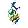
THIS ENTRY 3F6U REFLECTS AN ALTERNATIVE MODELING OF THE ORIGINAL STRUCTURAL DATA R1AUTSF DETERMINED ...THIS ENTRY 3F6U REFLECTS AN ALTERNATIVE MODELING OF THE ORIGINAL STRUCTURAL DATA R1AUTSF DETERMINED BY AUTHORS OF THE PDB ENTRY 1AUT: AUTHOR T.MATHER,V.OGANESSYAN,P.HOF,W.BODE,R.HUBER,S.FOUNDLING,C.ESMON
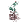
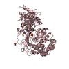
Vitamin K-dependent protein C ... , 2 types, 2 molecules HL
#1: Protein
VitaminK-dependentproteinCheavychain / Autoprothrombin IIA / Anticoagulant protein C / Blood coagulation factor XIV
Mass: 26965.836 Da / Num. of mol.: 1 / Fragment: UNP residues 212-451 / Source method: isolated from a natural source / Source: (natural) Homo sapiens (human) / References: UniProt: P04070, protein C (activated)
#2: Protein
VitaminK-dependentproteinClightchain / Autoprothrombin IIA / Anticoagulant protein C / Blood coagulation factor XIV
Mass: 10802.432 Da / Num. of mol.: 1 / Fragment: UNP residues 91-188 / Source method: isolated from a natural source / Source: (natural) Homo sapiens (human) / References: UniProt: P04070, protein C (activated)
Mass: 18.015 Da / Num. of mol.: 147 / Source method: isolated from a natural source / Formula: H2O
-
Details
Nonpolymer details
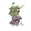
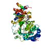
THE INHIBITOR IS BOUND TO THE ACTIVE SITE OF THE ENZYME. THE UNBOUND FORM OF THE INHIBITOR IS D-PHE- ...THE INHIBITOR IS BOUND TO THE ACTIVE SITE OF THE ENZYME. THE UNBOUND FORM OF THE INHIBITOR IS D-PHE-PRO-ARG-CHLOROMETHYLKETONE. UPON REACTION WITH PROTEIN IT FORMS TWO COVALENT BONDS: 1) A COVALENT BOND TO SER 195 FORMING A HEMIKETAL AR7 AND 2) A COVALENT BOND TO NE2 OF HIS 57
-
Experimental details
-
Experiment
Experiment
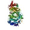
Method: X-RAY DIFFRACTION
-
Sample preparation
Crystal
Density Matthews: 3.43 Å3/Da / Density % sol: 64.1 % / Description: AUTHORS USED THE SF DATA FROM ENTRY 1AUT.
In the structure databanks used in Yorodumi, some data are registered as the other names, "COVID-19 virus" and "2019-nCoV". Here are the details of the virus and the list of structure data.
Jan 31, 2019. EMDB accession codes are about to change! (news from PDBe EMDB page)
EMDB accession codes are about to change! (news from PDBe EMDB page)
The allocation of 4 digits for EMDB accession codes will soon come to an end. Whilst these codes will remain in use, new EMDB accession codes will include an additional digit and will expand incrementally as the available range of codes is exhausted. The current 4-digit format prefixed with “EMD-” (i.e. EMD-XXXX) will advance to a 5-digit format (i.e. EMD-XXXXX), and so on. It is currently estimated that the 4-digit codes will be depleted around Spring 2019, at which point the 5-digit format will come into force.
The EM Navigator/Yorodumi systems omit the EMD- prefix.
Related info.:Q: What is EMD? / ID/Accession-code notation in Yorodumi/EM Navigator
Yorodumi is a browser for structure data from EMDB, PDB, SASBDB, etc.
This page is also the successor to EM Navigator detail page, and also detail information page/front-end page for Omokage search.
The word "yorodu" (or yorozu) is an old Japanese word meaning "ten thousand". "mi" (miru) is to see.
Related info.:EMDB / PDB / SASBDB / Comparison of 3 databanks / Yorodumi Search / Aug 31, 2016. New EM Navigator & Yorodumi / Yorodumi Papers / Jmol/JSmol / Function and homology information / Changes in new EM Navigator and Yorodumi
 Movie
Movie Controller

 Yorodumi
 Open data
 Basic information
 Components
 Keywords
 BLOOD COAGULATION /
  Function and homology information
 Authors
 Citation
 Structure visualization
 Downloads & links
 Other downloads

 PDBj
PDBj


 Assembly


 Type: peptide-like
 Mass: 22.990 Da / Num. of mol.: 1 / Source method: obtained synthetically / Formula: Na
 Mass: 40.078 Da / Num. of mol.: 1 / Source method: obtained synthetically / Formula: Ca
 Sample preparation
 Processing