 Movie
Movie Controller

[English] 日本語
 Yorodumi
- PDB-3f3b: Structure of the phage-like element PBSX protein xkdH from Bacill... -
+ Open data

- Basic information
| Entry | Database: PDB / ID: 3f3b | ||||||
|---|---|---|---|---|---|---|---|
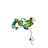
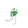
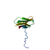
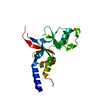
| Title | Structure of the phage-like element PBSX protein xkdH from Bacillus Subtilus. Northeast Structural Genomics Consortium target SR352. | ||||||
 Components Components | Phage-like element PBSX protein xkdH | ||||||
 Keywords Keywords |  structural genomics / unknown function / NESG X-Ray SR362 P54328 Structure / PSI-2 / Protein Structure Initiative / Northeast Structural Genomics Consortium structural genomics / unknown function / NESG X-Ray SR362 P54328 Structure / PSI-2 / Protein Structure Initiative / Northeast Structural Genomics Consortium | ||||||
| Function / homology | Protein of unknown function DUF3599 / Protein of unknown function DUF3599 / XkdH-like superfamily / Domain of unknown function (DUF3599) / Thrombin, subunit H / Beta Barrel / Mainly Beta / PHOSPHATE ION / Phage-like element PBSX protein XkdH Function and homology information Function and homology information | ||||||
| Biological species |  Bacillus subtilis (bacteria) Bacillus subtilis (bacteria) | ||||||
| Method | X-RAY DIFFRACTION / SYNCHROTRON / SAD / Resolution: 2.5 Å | ||||||
 Authors Authors | Kuzin, A.P. / Neely, H. / Seetharaman, J. / Clayton, G. / Ho, C.K. / Janjua, H. / Cunningham, K. / Ma, L.-C. / Xiao, R. / Liu, J. ...Kuzin, A.P. / Neely, H. / Seetharaman, J. / Clayton, G. / Ho, C.K. / Janjua, H. / Cunningham, K. / Ma, L.-C. / Xiao, R. / Liu, J. / Baran, M.C. / Acton, T.B. / Rost, B. / Montelione, G.T. / Tong, L. / Hunt, J.F. / Northeast Structural Genomics Consortium (NESG) | ||||||
 Citation Citation | Journal: To be Published Title: Structure of the phage-like element PBSX protein xkdH from Bacillus Subtilus. Northeast Structural Genomics Consortium target SR352. Authors: Kuzin, A.P. / Neely, H. / Seetharaman, J. / Clayton, G. / Ho, C.K. / Cunningham, K. / Ma, L.-C. / Xiao, R. / Liu, J. / Baran, M.C. / Acton, T.B. / Rost, B. / Montelione, G.T. / Tong, L. / Hunt, J.F. | ||||||
| History |
|
- Structure visualization
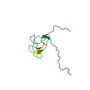
| Structure viewer | Molecule: MolmilJmol/JSmol |
|---|
- Downloads & links
-Download
| PDBx/mmCIF format | 3f3b.cif.gz | 38.3 KB | Display | PDBx/mmCIF format |
|---|---|---|---|---|
| PDB format | pdb3f3b.ent.gz | 26.2 KB | Display | PDB format |
| PDBx/mmJSON format | 3f3b.json.gz | Tree view | PDBx/mmJSON format | |
| Others |  Other downloads Other downloads |
-Validation report
| Arichive directory | https://data.pdbj.org/pub/pdb/validation_reports/f3/3f3bftp://data.pdbj.org/pub/pdb/validation_reports/f3/3f3b | HTTPS FTP |
|---|
-Related structure data
| Similar structure data | |
|---|---|
| Other databases |


-Links
 PDBj
PDBj- Assembly
| Deposited unit | 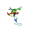
| ||||||||
|---|---|---|---|---|---|---|---|---|---|
| 1 |
| ||||||||
| Unit cell |
|
-Components
| #1: Protein | Mass: 15164.803 Da / Num. of mol.: 1 Source method: isolated from a genetically manipulated source Source: (gene. exp.) Bacillus subtilis (bacteria) / Gene: xkdH, BSU12620 / References: UniProt: P54328 | ||
|---|---|---|---|
| #2: Chemical | Phosphate  Mass: 94.971 Da / Num. of mol.: 2 / Source method: obtained synthetically / Formula: PO4 Mass: 94.971 Da / Num. of mol.: 2 / Source method: obtained synthetically / Formula: PO4#3: Water | ChemComp-HOH / | Water Mass: 18.015 Da / Num. of mol.: 60 / Source method: isolated from a natural source / Formula: H2O Mass: 18.015 Da / Num. of mol.: 60 / Source method: isolated from a natural source / Formula: H2O |
-Experimental details
-Experiment
| Experiment | Method: X-RAY DIFFRACTION / Number of used crystals: 1 |
|---|
- Sample preparation
| Crystal | Density Matthews: 2.46 Å3/Da / Density % sol: 49.93 % |
|---|---|
| Crystal grow | Temperature: 277 K / Method: vapor diffusion, hanging drop / pH: 7.5 Details: 0.55M NaH2PO4, 0.5M KH2PO4, 0.08M Hepes-Na, pH 7.5, VAPOR DIFFUSION, HANGING DROP, temperature 277K |
-Data collection
| Diffraction | Mean temperature: 100 K |
|---|---|
| Diffraction source | Source: SYNCHROTRON / Site: NSLS  / Beamline: X4A / Wavelength: 0.979 / Beamline: X4A / Wavelength: 0.979 |
| Detector | Type: ADSC QUANTUM 4 / Detector: CCD / Date: Aug 14, 2008 / Details: mirrors |
| Radiation | Protocol: SINGLE WAVELENGTH / Monochromatic (M) / Laue (L): M / Scattering type: x-ray |
| Radiation wavelength | Wavelength: 0.979 Å / Relative weight: 1 |
| Reflection | Resolution: 2.5→30 Å / Num. obs: 9878 / % possible obs: 98.3 % / Observed criterion σ(I): -3 / Redundancy: 10 % / Biso Wilson estimate: 41.2 Å2 / Rmerge(I) obs: 0.047 / Net I/σ(I): 49.3 |
| Reflection shell | Resolution: 2.5→2.59 Å / Redundancy: 6 % / Rmerge(I) obs: 0.117 / Mean I/σ(I) obs: 16.9 / Num. unique all: 922 / % possible all: 96.4 |
- Processing
| Software |
| ||||||||||||||||||||||||||||||||||||
|---|---|---|---|---|---|---|---|---|---|---|---|---|---|---|---|---|---|---|---|---|---|---|---|---|---|---|---|---|---|---|---|---|---|---|---|---|---|
| Refinement | Method to determine structure: SAD / Resolution: 2.5→19.97 Å / Rfactor Rfree error: 0.012 / Data cutoff high absF: 268301.63 / Data cutoff low absF: 0 / Isotropic thermal model: RESTRAINED / Cross valid method: THROUGHOUT / σ(F): 1 / Stereochemistry target values: Engh & Huber / Details: BULK SOLVENT MODEL USED
| ||||||||||||||||||||||||||||||||||||
| Solvent computation | Solvent model: FLAT MODEL / Bsol: 37.2987 Å2 / ksol: 0.4 e/Å3 | ||||||||||||||||||||||||||||||||||||
| Displacement parameters | Biso mean: 36.3 Å2
| ||||||||||||||||||||||||||||||||||||
| Refine analyze |
| ||||||||||||||||||||||||||||||||||||
| Refinement step | Cycle: LAST / Resolution: 2.5→19.97 Å
| ||||||||||||||||||||||||||||||||||||
| Refine LS restraints |
| ||||||||||||||||||||||||||||||||||||
| LS refinement shell | Resolution: 2.5→2.66 Å / Rfactor Rfree error: 0.032 / Total num. of bins used: 6
| ||||||||||||||||||||||||||||||||||||
| Xplor file |
|
