 Movie
Movie Controller

[English] 日本語
 Yorodumi
- PDB-3ew0: The novel 2Fe-2S outer mitochondrial protein mitoNEET displays co... -
+ Open data

- Basic information
| Entry | Database: PDB / ID: 3ew0 | ||||||
|---|---|---|---|---|---|---|---|
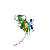
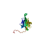
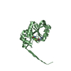
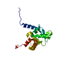
| Title | The novel 2Fe-2S outer mitochondrial protein mitoNEET displays conformational flexibility in its N-terminal cytoplasmic tethering domain | ||||||
 Components Components | CDGSH iron sulfur domain-containing protein 1 | ||||||
 Keywords Keywords | METAL BINDING PROTEIN / mitochondrial outer membrane / 2Fe-2S proteins / isotopic labeling / highyield expression /  Iron / Iron-sulfur / Membrane / Metal-binding / Mitochondrion / Mitochondrion outer membrane / Signal-anchor / Transmembrane Iron / Iron-sulfur / Membrane / Metal-binding / Mitochondrion / Mitochondrion outer membrane / Signal-anchor / Transmembrane | ||||||
| Function / homology |  Function and homology information Function and homology informationprotein maturation by [2Fe-2S] cluster transfer / cysteine transaminase / L-cysteine transaminase activity / regulation of cellular respiration / regulation of autophagy / 2 iron, 2 sulfur cluster binding / pyridoxal phosphate binding / mitochondrial outer membrane / protein homodimerization activity / mitochondrion ...protein maturation by [2Fe-2S] cluster transfer / cysteine transaminase / L-cysteine transaminase activity / regulation of cellular respiration / regulation of autophagy / 2 iron, 2 sulfur cluster binding / pyridoxal phosphate binding / mitochondrial outer membrane / protein homodimerization activity / mitochondrion / identical protein binding / metal ion bindingSimilarity search - Function | ||||||
| Biological species |  Homo sapiens (human) Homo sapiens (human) | ||||||
| Method | X-RAY DIFFRACTION / SYNCHROTRON / MOLECULAR REPLACEMENT / Resolution: 1.4 Å | ||||||
 Authors Authors | Conlan, A.R. / Paddock, M.L. / Wiley, S. / Axelrod, H.L. / Cohen, A.E. / Abresch, E.C. / Roy, M. / Nechushtai, R. / Jennings, P.A. | ||||||
 Citation Citation | Journal: Acta Crystallogr.,Sect.F / Year: 2009 Title: The novel 2Fe-2S outer mitochondrial protein mitoNEET displays conformational flexibility in its N-terminal cytoplasmic tethering domain. Authors: Conlan, A.R. / Paddock, M.L. / Axelrod, H.L. / Cohen, A.E. / Abresch, E.C. / Wiley, S. / Roy, M. / Nechushtai, R. / Jennings, P.A. | ||||||
| History |
|
- Structure visualization
| Structure viewer | Molecule: MolmilJmol/JSmol |
|---|
- Downloads & links
-Download
| PDBx/mmCIF format | 3ew0.cif.gz | 49.4 KB | Display | PDBx/mmCIF format |
|---|---|---|---|---|
| PDB format | pdb3ew0.ent.gz | 34.3 KB | Display | PDB format |
| PDBx/mmJSON format | 3ew0.json.gz | Tree view | PDBx/mmJSON format | |
| Others |  Other downloads Other downloads |
-Validation report
| Arichive directory | https://data.pdbj.org/pub/pdb/validation_reports/ew/3ew0ftp://data.pdbj.org/pub/pdb/validation_reports/ew/3ew0 | HTTPS FTP |
|---|
-Related structure data
| Related structure data |  2qh7S S: Starting model for refinement |
|---|---|
| Similar structure data |


-Links
 PDBj
PDBj


- Assembly
| Deposited unit | 
| ||||||||
|---|---|---|---|---|---|---|---|---|---|
| 1 |
| ||||||||
| Unit cell |
|
-Components
| #1: Protein | Mass: 9280.703 Da / Num. of mol.: 2 / Fragment: UNP residues 33-108, WATER-SOLULE DOMAIN Source method: isolated from a genetically manipulated source Source: (gene. exp.) Homo sapiens (human) / Gene: C10orf70, CISD1, MDS029, ZCD1Plasmid: modified pET28-a(+) vector (Novagen) that contained the superfolder GFP cDNA Production host:  Escherichia coli (E. coli) / Strain (production host): E. coli BL-21(DE3/pET-24d) / References: UniProt: Q9NZ45 Escherichia coli (E. coli) / Strain (production host): E. coli BL-21(DE3/pET-24d) / References: UniProt: Q9NZ45#2: Chemical | Iron–sulfur cluster  Mass: 175.820 Da / Num. of mol.: 2 / Source method: obtained synthetically / Formula: Fe2S2 Mass: 175.820 Da / Num. of mol.: 2 / Source method: obtained synthetically / Formula: Fe2S2#3: Water | ChemComp-HOH / | Water Mass: 18.015 Da / Num. of mol.: 164 / Source method: isolated from a natural source / Formula: H2O Mass: 18.015 Da / Num. of mol.: 164 / Source method: isolated from a natural source / Formula: H2O |
|---|
-Experimental details
-Experiment
| Experiment | Method: X-RAY DIFFRACTION / Number of used crystals: 1 |
|---|
- Sample preparation
| Crystal | Density Matthews: 1.96 Å3/Da / Density % sol: 37.33 % |
|---|---|
| Crystal grow | Temperature: 300 K / Method: vapor diffusion / pH: 8 Details: Crystallization Droplets: 100 mM Tris HCl (pH 8.0), 100 mM NaCl, and 16-18% PEG 3000. Reservoir conditions: 100 mM Tris-HCl (pH 8.0), 100 mM NaCl, and 30 32% PEG 3000 in the reservoir., VAPOR DIFFUSION |
-Data collection
| Diffraction | Mean temperature: 100 K |
|---|---|
| Diffraction source | Source: SYNCHROTRON / Site: ALS  / Beamline: 12.3.1 / Wavelength: 1.1158 Å / Beamline: 12.3.1 / Wavelength: 1.1158 Å |
| Detector | Type: ADSC QUANTUM 315 / Detector: CCD / Date: Oct 12, 2007 / Details: Kohzu Dual Double Crystal Monochromator (DDCM) |
| Radiation | Monochromator: double Si(111) crystals / Protocol: SINGLE WAVELENGTH / Monochromatic (M) / Laue (L): M / Scattering type: x-ray |
| Radiation wavelength | Wavelength: 1.1158 Å / Relative weight: 1 |
| Reflection | Resolution: 1.4→38.55 Å / Num. all: 26874 / Num. obs: 26874 / % possible obs: 92.4 % / Observed criterion σ(I): 0 / Redundancy: 6.3 % / Rsym value: 0.059 / Net I/σ(I): 19 |
| Reflection shell | Resolution: 1.4→1.48 Å / Redundancy: 4.6 % / Mean I/σ(I) obs: 4.5 / Rsym value: 0.32 / % possible all: 81.4 |
- Processing
| Software |
| |||||||||||||||||||||||||||||||||||||||||||||||||||||||||||||||||||||||||||||||||||||||||||||||||||||||||||||||||||||||||||||||||||||||||||||||||||||||||||||||||||||||||||||||
|---|---|---|---|---|---|---|---|---|---|---|---|---|---|---|---|---|---|---|---|---|---|---|---|---|---|---|---|---|---|---|---|---|---|---|---|---|---|---|---|---|---|---|---|---|---|---|---|---|---|---|---|---|---|---|---|---|---|---|---|---|---|---|---|---|---|---|---|---|---|---|---|---|---|---|---|---|---|---|---|---|---|---|---|---|---|---|---|---|---|---|---|---|---|---|---|---|---|---|---|---|---|---|---|---|---|---|---|---|---|---|---|---|---|---|---|---|---|---|---|---|---|---|---|---|---|---|---|---|---|---|---|---|---|---|---|---|---|---|---|---|---|---|---|---|---|---|---|---|---|---|---|---|---|---|---|---|---|---|---|---|---|---|---|---|---|---|---|---|---|---|---|---|---|---|---|---|
| Refinement | Method to determine structure: MOLECULAR REPLACEMENT Starting model: PDB ENTRY 2QH7 Resolution: 1.4→38.55 Å / Cor.coef. Fo:Fc: 0.941 / Cor.coef. Fo:Fc free: 0.912 / SU B: 2.027 / SU ML: 0.051 / TLS residual ADP flag: LIKELY RESIDUAL / Cross valid method: THROUGHOUT / ESU R: 0.077 / ESU R Free: 0.082 / Stereochemistry target values: MAXIMUM LIKELIHOOD Details: 1. HYDROGENS HAVE BEEN ADDED IN THE RIDING POSITIONS. 2. ATOM RECORD CONTAINS RESIDUAL B FACTORS ONLY.
| |||||||||||||||||||||||||||||||||||||||||||||||||||||||||||||||||||||||||||||||||||||||||||||||||||||||||||||||||||||||||||||||||||||||||||||||||||||||||||||||||||||||||||||||
| Solvent computation | Ion probe radii: 0.8 Å / Shrinkage radii: 0.8 Å / VDW probe radii: 1.2 Å / Solvent model: BABINET MODEL WITH MASK | |||||||||||||||||||||||||||||||||||||||||||||||||||||||||||||||||||||||||||||||||||||||||||||||||||||||||||||||||||||||||||||||||||||||||||||||||||||||||||||||||||||||||||||||
| Displacement parameters | Biso mean: 5.626 Å2
| |||||||||||||||||||||||||||||||||||||||||||||||||||||||||||||||||||||||||||||||||||||||||||||||||||||||||||||||||||||||||||||||||||||||||||||||||||||||||||||||||||||||||||||||
| Refinement step | Cycle: LAST / Resolution: 1.4→38.55 Å
| |||||||||||||||||||||||||||||||||||||||||||||||||||||||||||||||||||||||||||||||||||||||||||||||||||||||||||||||||||||||||||||||||||||||||||||||||||||||||||||||||||||||||||||||
| Refine LS restraints |
| |||||||||||||||||||||||||||||||||||||||||||||||||||||||||||||||||||||||||||||||||||||||||||||||||||||||||||||||||||||||||||||||||||||||||||||||||||||||||||||||||||||||||||||||
| LS refinement shell | Resolution: 1.401→1.437 Å / Total num. of bins used: 20
| |||||||||||||||||||||||||||||||||||||||||||||||||||||||||||||||||||||||||||||||||||||||||||||||||||||||||||||||||||||||||||||||||||||||||||||||||||||||||||||||||||||||||||||||
| Refinement TLS params. | Method: refined / Refine-ID: X-RAY DIFFRACTION
| |||||||||||||||||||||||||||||||||||||||||||||||||||||||||||||||||||||||||||||||||||||||||||||||||||||||||||||||||||||||||||||||||||||||||||||||||||||||||||||||||||||||||||||||
| Refinement TLS group |
|
