 Movie
Movie Controller

[English] 日本語
 Yorodumi
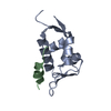
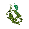
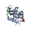
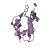
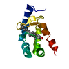
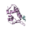
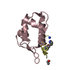
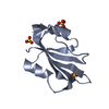
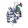
- PDB-3eqy: Crystal structure of human MDMX in complex with a 12-mer peptide ... -
+ Open data

- Basic information
| Entry | Database: PDB / ID: 3eqy | ||||||
|---|---|---|---|---|---|---|---|
| Title | Crystal structure of human MDMX in complex with a 12-mer peptide inhibitor | ||||||
 Components Components |
| ||||||
 Keywords Keywords |  ONCOPROTEIN / MDM4 / MDMX / MDMX-peptide inhibitor complex / Metal-binding / Nucleus / Zinc-finger ONCOPROTEIN / MDM4 / MDMX / MDMX-peptide inhibitor complex / Metal-binding / Nucleus / Zinc-finger | ||||||
| Function / homology |  Function and homology information Function and homology informationatrial septum development / heart valve development / atrioventricular valve morphogenesis / ventricular septum development / endocardial cushion morphogenesis / DNA damage response, signal transduction by p53 class mediator / transcription repressor complex / Stabilization of p53 / Oncogene Induced Senescence / negative regulation of protein catabolic process ...atrial septum development / heart valve development / atrioventricular valve morphogenesis / ventricular septum development / endocardial cushion morphogenesis / DNA damage response, signal transduction by p53 class mediator / transcription repressor complex / Stabilization of p53 / Oncogene Induced Senescence / negative regulation of protein catabolic process / Regulation of TP53 Activity through Methylation / ubiquitin-protein transferase activity / Regulation of TP53 Degradation / cellular response to hypoxia / protein-containing complex assembly / Oxidative Stress Induced Senescence / Regulation of TP53 Activity through Phosphorylation / protein stabilization / Ub-specific processing proteases / protein ubiquitination / regulation of cell cycle / negative regulation of cell population proliferation / negative regulation of DNA-templated transcription / negative regulation of apoptotic process / enzyme binding / negative regulation of transcription by RNA polymerase II / zinc ion binding / nucleoplasm / nucleusSimilarity search - Function | ||||||
| Biological species |  Homo sapiens (human) Homo sapiens (human) | ||||||
| Method | X-RAY DIFFRACTION / MOLECULAR REPLACEMENT / Resolution: 1.63 Å | ||||||
 Authors Authors | Pazgier, M. / Lu, W. | ||||||
 Citation Citation | Journal: Proc.Natl.Acad.Sci.USA / Year: 2009 Title: Structural basis for high-affinity peptide inhibition of p53 interactions with MDM2 and MDMX. Authors: Pazgier, M. / Liu, M. / Zou, G. / Yuan, W. / Li, C. / Li, C. / Li, J. / Monbo, J. / Zella, D. / Tarasov, S.G. / Lu, W. | ||||||
| History |
|
- Structure visualization
| Structure viewer | Molecule: MolmilJmol/JSmol |
|---|
- Downloads & links
-Download
| PDBx/mmCIF format | 3eqy.cif.gz | 60.3 KB | Display | PDBx/mmCIF format |
|---|---|---|---|---|
| PDB format | pdb3eqy.ent.gz | 44.2 KB | Display | PDB format |
| PDBx/mmJSON format | 3eqy.json.gz | Tree view | PDBx/mmJSON format | |
| Others |  Other downloads Other downloads |
-Validation report
| Arichive directory | https://data.pdbj.org/pub/pdb/validation_reports/eq/3eqyftp://data.pdbj.org/pub/pdb/validation_reports/eq/3eqy | HTTPS FTP |
|---|
-Related structure data
| Related structure data |  3eqsC 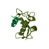 2z5tS C: citing same article ( S: Starting model for refinement |
|---|---|
| Similar structure data |


-Links
 PDBj
PDBj

- Assembly
| Deposited unit | 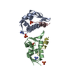
| ||||||||||||||||||
|---|---|---|---|---|---|---|---|---|---|---|---|---|---|---|---|---|---|---|---|
| 1 | 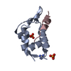
| ||||||||||||||||||
| 2 | 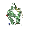
| ||||||||||||||||||
| Unit cell |
| ||||||||||||||||||
| Noncrystallographic symmetry (NCS) | NCS domain:
|
-Components
| #1: Protein | / p53-binding protein Mdm4 / Mdm2-like p53-binding protein / Protein Mdmx / Double minute 4 protein Mass: 9575.293 Da / Num. of mol.: 2 / Fragment: UNP residues residues 24-108 / Mutation: Q68A, Q69A, E70A / Source method: obtained synthetically / Source: (synth.) Homo sapiens (human) / References: UniProt: O15151#2: Protein/peptide | Mass: 1427.557 Da / Num. of mol.: 2 / Source method: obtained synthetically Details: peptide identified by screening a duodecimal peptide library displayed on M13 phage #3: Chemical | Guanidine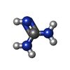  Mass: 59.070 Da / Num. of mol.: 2 / Source method: obtained synthetically / Formula: CH5N3 Mass: 59.070 Da / Num. of mol.: 2 / Source method: obtained synthetically / Formula: CH5N3#4: Chemical | ChemComp-PO4 / Phosphate  Mass: 94.971 Da / Num. of mol.: 4 / Source method: obtained synthetically / Formula: PO4 Mass: 94.971 Da / Num. of mol.: 4 / Source method: obtained synthetically / Formula: PO4#5: Water | ChemComp-HOH / | Water Mass: 18.015 Da / Num. of mol.: 260 / Source method: isolated from a natural source / Formula: H2O Mass: 18.015 Da / Num. of mol.: 260 / Source method: isolated from a natural source / Formula: H2O |
|---|
-Experimental details
-Experiment
| Experiment | Method: X-RAY DIFFRACTION / Number of used crystals: 1 |
|---|
- Sample preparation
| Crystal | Density Matthews: 2.64 Å3/Da / Density % sol: 53.4 % |
|---|---|
| Crystal grow | Temperature: 298 K / Method: vapor diffusion, hanging drop / pH: 7.5 Details: 0.1M HEPES, 0.8 M sodium phosphate, 0.8 M potassium phosphate, pH 7.5, VAPOR DIFFUSION, HANGING DROP, temperature 298K |
-Data collection
| Diffraction | Mean temperature: 100 K |
|---|---|
| Diffraction source | Source: ROTATING ANODE / Type: RIGAKU MICROMAX-007 / Wavelength: 1.54 Å |
| Detector | Type: RIGAKU RAXIS IV++ / Detector: IMAGE PLATE / Date: May 27, 2008 / Details: confocal |
| Radiation | Protocol: SINGLE WAVELENGTH / Monochromatic (M) / Laue (L): M / Scattering type: x-ray |
| Radiation wavelength | Wavelength: 1.54 Å / Relative weight: 1 |
| Reflection | Resolution: 1.63→65.51 Å / Num. all: 27997 / Num. obs: 27972 / % possible obs: 99.9 % / Redundancy: 5.2 % / Rmerge(I) obs: 0.045 / Rsym value: 0.049 / Net I/σ(I): 64.3 |
| Reflection shell | Resolution: 1.63→1.69 Å / Redundancy: 4.8 % / Rmerge(I) obs: 0.101 / Mean I/σ(I) obs: 20.2 / Rsym value: 0.098 / % possible all: 94.8 |
- Processing
| Software |
| ||||||||||||||||||||||||||||||||||||||||||||||||||||||||||||||||||||||||||||||||||||||||||||||||||||||||||||||||||||||||||||||||||||||||||||||||||||||||||||||||||||||||||
|---|---|---|---|---|---|---|---|---|---|---|---|---|---|---|---|---|---|---|---|---|---|---|---|---|---|---|---|---|---|---|---|---|---|---|---|---|---|---|---|---|---|---|---|---|---|---|---|---|---|---|---|---|---|---|---|---|---|---|---|---|---|---|---|---|---|---|---|---|---|---|---|---|---|---|---|---|---|---|---|---|---|---|---|---|---|---|---|---|---|---|---|---|---|---|---|---|---|---|---|---|---|---|---|---|---|---|---|---|---|---|---|---|---|---|---|---|---|---|---|---|---|---|---|---|---|---|---|---|---|---|---|---|---|---|---|---|---|---|---|---|---|---|---|---|---|---|---|---|---|---|---|---|---|---|---|---|---|---|---|---|---|---|---|---|---|---|---|---|---|---|---|
| Refinement | Method to determine structure: MOLECULAR REPLACEMENT Starting model: PDB entry 2Z5T Resolution: 1.63→15 Å / Cor.coef. Fo:Fc: 0.971 / Cor.coef. Fo:Fc free: 0.965 / SU B: 2.5 / SU ML: 0.041 / TLS residual ADP flag: LIKELY RESIDUAL / Cross valid method: THROUGHOUT / ESU R: 0.082 / ESU R Free: 0.075 / Stereochemistry target values: MAXIMUM LIKELIHOOD / Details: HYDROGENS HAVE BEEN ADDED IN THE RIDING POSITIONS
| ||||||||||||||||||||||||||||||||||||||||||||||||||||||||||||||||||||||||||||||||||||||||||||||||||||||||||||||||||||||||||||||||||||||||||||||||||||||||||||||||||||||||||
| Solvent computation | Ion probe radii: 0.8 Å / Shrinkage radii: 0.8 Å / VDW probe radii: 1.2 Å / Solvent model: BABINET MODEL WITH MASK | ||||||||||||||||||||||||||||||||||||||||||||||||||||||||||||||||||||||||||||||||||||||||||||||||||||||||||||||||||||||||||||||||||||||||||||||||||||||||||||||||||||||||||
| Displacement parameters | Biso mean: 15.887 Å2
| ||||||||||||||||||||||||||||||||||||||||||||||||||||||||||||||||||||||||||||||||||||||||||||||||||||||||||||||||||||||||||||||||||||||||||||||||||||||||||||||||||||||||||
| Refinement step | Cycle: LAST / Resolution: 1.63→15 Å
| ||||||||||||||||||||||||||||||||||||||||||||||||||||||||||||||||||||||||||||||||||||||||||||||||||||||||||||||||||||||||||||||||||||||||||||||||||||||||||||||||||||||||||
| Refine LS restraints |
| ||||||||||||||||||||||||||||||||||||||||||||||||||||||||||||||||||||||||||||||||||||||||||||||||||||||||||||||||||||||||||||||||||||||||||||||||||||||||||||||||||||||||||
| Refine LS restraints NCS | Refine-ID: X-RAY DIFFRACTION
| ||||||||||||||||||||||||||||||||||||||||||||||||||||||||||||||||||||||||||||||||||||||||||||||||||||||||||||||||||||||||||||||||||||||||||||||||||||||||||||||||||||||||||
| LS refinement shell | Resolution: 1.63→1.674 Å / Total num. of bins used: 20
| ||||||||||||||||||||||||||||||||||||||||||||||||||||||||||||||||||||||||||||||||||||||||||||||||||||||||||||||||||||||||||||||||||||||||||||||||||||||||||||||||||||||||||
| Refinement TLS params. | Method: refined / Refine-ID: X-RAY DIFFRACTION
| ||||||||||||||||||||||||||||||||||||||||||||||||||||||||||||||||||||||||||||||||||||||||||||||||||||||||||||||||||||||||||||||||||||||||||||||||||||||||||||||||||||||||||
| Refinement TLS group |
|
