Resolution: 1.9→50 Å / Cross valid method: THROUGHOUT / σ(F): 0 / σ(I): 0 / Stereochemistry target values: MAXIMUM LIKELYHOOD / Details: HYDROGENS HAVE BEEN ADDED IN THE RIDING POSITIONS
Rfactor
Num. reflection
% reflection
Selection details
Rfree
0.22913
1762
5.3 %
RANDOM
Rwork
0.18132
-
-
-
all
0.18374
33439
-
-
obs
0.18374
33439
96.54 %
-
Displacement parameters
Biso mean: 17.86 Å2
Baniso -1
Baniso -2
Baniso -3
1-
0.41 Å2
0 Å2
0 Å2
2-
-
-1.73 Å2
0 Å2
3-
-
-
1.32 Å2
Refinement step
Cycle: LAST / Resolution: 1.9→50 Å
Protein
Nucleic acid
Ligand
Solvent
Total
Num. atoms
3474
0
235
390
4099
Refine LS restraints
Refine-ID
Type
Dev ideal
X-RAY DIFFRACTION
r_bond_refined_d
0.009
X-RAY DIFFRACTION
r_angle_refined_deg
1.15
X-RAY DIFFRACTION
r_chiral_restr
0.076
X-RAY DIFFRACTION
r_gen_planes_refined
0.004
X-RAY DIFFRACTION
r_nbd_refined
0.186
X-RAY DIFFRACTION
r_nbtor_refined
0.304
X-RAY DIFFRACTION
r_symmetry_vdw_refined
0.119
X-RAY DIFFRACTION
r_symmetry_hbond_refined
0.131
LS refinement shell
Resolution: 1.9→1.949 Å
Rfactor
Num. reflection
% reflection
Rfree
0.277
67
-
Rwork
0.233
-
-
obs
-
1720
67.72 %
+
About Yorodumi
-
News
-
Feb 9, 2022. New format data for meta-information of EMDB entries
New format data for meta-information of EMDB entries
Version 3 of the EMDB header file is now the official format.
The previous official version 1.9 will be removed from the archive.
In the structure databanks used in Yorodumi, some data are registered as the other names, "COVID-19 virus" and "2019-nCoV". Here are the details of the virus and the list of structure data.
Jan 31, 2019. EMDB accession codes are about to change! (news from PDBe EMDB page)
EMDB accession codes are about to change! (news from PDBe EMDB page)
The allocation of 4 digits for EMDB accession codes will soon come to an end. Whilst these codes will remain in use, new EMDB accession codes will include an additional digit and will expand incrementally as the available range of codes is exhausted. The current 4-digit format prefixed with “EMD-” (i.e. EMD-XXXX) will advance to a 5-digit format (i.e. EMD-XXXXX), and so on. It is currently estimated that the 4-digit codes will be depleted around Spring 2019, at which point the 5-digit format will come into force.
The EM Navigator/Yorodumi systems omit the EMD- prefix.
Related info.:Q: What is EMD? / ID/Accession-code notation in Yorodumi/EM Navigator
Yorodumi is a browser for structure data from EMDB, PDB, SASBDB, etc.
This page is also the successor to EM Navigator detail page, and also detail information page/front-end page for Omokage search.
The word "yorodu" (or yorozu) is an old Japanese word meaning "ten thousand". "mi" (miru) is to see.
Related info.:EMDB / PDB / SASBDB / Comparison of 3 databanks / Yorodumi Search / Aug 31, 2016. New EM Navigator & Yorodumi / Yorodumi Papers / Jmol/JSmol / Function and homology information / Changes in new EM Navigator and Yorodumi
 Movie
Movie Controller

 Yorodumi
 Open data
 Basic information
 Components
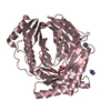
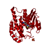
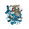
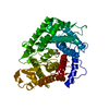
 Glucan 1,4-a-glucosidase
  Keywords
 Function and homology information
 Authors
 Citation
 Structure visualization
 Downloads & links
 Other downloads
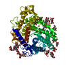


 PDBj
PDBj


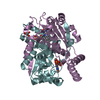
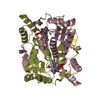
 Assembly
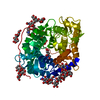

 Type: D-saccharide, alpha linking / Mass: 180.156 Da / Num. of mol.: 7


 Mass: 122.143 Da / Num. of mol.: 1 / Source method: obtained synthetically / Formula: C4H12NO3 / Comment: pH buffer*YM
 Mass: 92.094 Da / Num. of mol.: 1 / Source method: obtained synthetically / Formula: C3H8O3
 Sample preparation
 / Beamline: 8.2.2 / Wavelength: 1 Å
 Processing