 Movie
Movie Controller

[English] 日本語
 Yorodumi
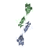
- PDB-3eoy: Structure of Reovirus sigma1 in Complex with Its Receptor Junctio... -
+ Open data

- Basic information
| Entry | Database: PDB / ID: 3eoy | ||||||
|---|---|---|---|---|---|---|---|
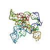
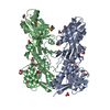
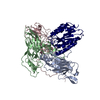
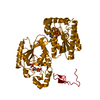
| Title | Structure of Reovirus sigma1 in Complex with Its Receptor Junctional Adhesion Molecule-A | ||||||
 Components Components |
| ||||||
 Keywords Keywords |  VIRAL PROTEIN/CELL ADHESION / protein complex / virus receptor complex / beta-barrel / beta-spiral repeat / greek key motif / trimer / immunoglobulin superfamily / VIRAL PROTEIN-CELL ADHESION COMPLEX VIRAL PROTEIN/CELL ADHESION / protein complex / virus receptor complex / beta-barrel / beta-spiral repeat / greek key motif / trimer / immunoglobulin superfamily / VIRAL PROTEIN-CELL ADHESION COMPLEX | ||||||
| Function / homology |  Function and homology information Function and homology informationpositive regulation of establishment of endothelial barrier / memory T cell extravasation / Tight junction interactions / establishment of endothelial intestinal barrier / regulation of membrane permeability / viral outer capsid / protein localization to bicellular tight junction / actomyosin structure organization / positive regulation of platelet aggregation / tight junction ...positive regulation of establishment of endothelial barrier / memory T cell extravasation / Tight junction interactions / establishment of endothelial intestinal barrier / regulation of membrane permeability / viral outer capsid / protein localization to bicellular tight junction / actomyosin structure organization / positive regulation of platelet aggregation / tight junction / regulation of bicellular tight junction assembly / intestinal absorption / negative regulation of stress fiber assembly / regulation of cytoskeleton organization / positive regulation of Rho protein signal transduction / leukocyte cell-cell adhesion / maintenance of blood-brain barrier / bicellular tight junction / Integrin cell surface interactions / regulation of cytokine production / TGF-beta receptor signaling in EMT (epithelial to mesenchymal transition) / PDZ domain binding / protein localization to plasma membrane / regulation of actin cytoskeleton organization / Cell surface interactions at the vascular wall / cell-cell adhesion / cellular response to mechanical stimulus / cell-cell junction / integrin binding / virus receptor activity / cell junction / regulation of cell shape / cell adhesion / cadherin binding / inflammatory response / symbiont entry into host cell / virion attachment to host cell / protein homodimerization activity / protein-containing complex / extracellular exosome / plasma membraneSimilarity search - Function | ||||||
| Biological species |  reovirus reovirus Homo sapiens (human) Homo sapiens (human) | ||||||
| Method | X-RAY DIFFRACTION / SYNCHROTRON / MOLECULAR REPLACEMENT / molecular replacement / Resolution: 3.4 Å | ||||||
 Authors Authors | Kirchner, E. / Guglielmi, K.M. / Dermody, T.S. / Stehle, T. | ||||||
 Citation Citation | Journal: Plos Pathog. / Year: 2008 Title: Structure of reovirus sigma1 in complex with its receptor junctional adhesion molecule-A Authors: Kirchner, E. / Guglielmi, K.M. / Strauss, H.M. / Dermody, T.S. / Stehle, T. | ||||||
| History |
|
- Structure visualization
| Structure viewer | Molecule: MolmilJmol/JSmol |
|---|
- Downloads & links
-Download
| PDBx/mmCIF format | 3eoy.cif.gz | 303.8 KB | Display | PDBx/mmCIF format |
|---|---|---|---|---|
| PDB format | pdb3eoy.ent.gz | 248.4 KB | Display | PDB format |
| PDBx/mmJSON format | 3eoy.json.gz | Tree view | PDBx/mmJSON format | |
| Others |  Other downloads Other downloads |
-Validation report
| Arichive directory | https://data.pdbj.org/pub/pdb/validation_reports/eo/3eoyftp://data.pdbj.org/pub/pdb/validation_reports/eo/3eoy | HTTPS FTP |
|---|
-Related structure data


-Links
 PDBj
PDBj


- Assembly
| Deposited unit | 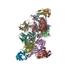
| ||||||||
|---|---|---|---|---|---|---|---|---|---|
| 1 | 
| ||||||||
| 2 | 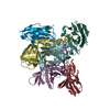
| ||||||||
| Unit cell |
|
-Components
| #1: Protein | Mass: 17921.033 Da / Num. of mol.: 6 / Fragment: head domain Source method: isolated from a genetically manipulated source Source: (gene. exp.) reovirus / Strain: TYPE 3/STRAIN DEARING / Gene: S1 / Plasmid: pGEX-4T-3 / Production host:  Escherichia coli (E. coli) / Strain (production host): BL21(DE3) pLysS / References: UniProt: P03528 Escherichia coli (E. coli) / Strain (production host): BL21(DE3) pLysS / References: UniProt: P03528#2: Protein | Mass: 11518.841 Da / Num. of mol.: 6 / Fragment: D1 domain Source method: isolated from a genetically manipulated source Source: (gene. exp.) Homo sapiens (human) / Plasmid: pGEX-4T-3 / Production host: Escherichia coli (E. coli) / Strain (production host): BL21(DE3) pLysS / References: UniProt: Q9Y624Sequence details | THIS SEQUENCE IS BASED ON REFERENCE 8 IN UNP DATABASE, P03528. | |
|---|
-Experimental details
-Experiment
| Experiment | Method: X-RAY DIFFRACTION / Number of used crystals: 3 |
|---|
- Sample preparation
| Crystal | Density Matthews: 2.44 Å3/Da / Density % sol: 49.51 % / Mosaicity: 1.39 ° |
|---|---|
| Crystal grow | Temperature: 293 K / Method: vapor diffusion, hanging drop / pH: 9.5 Details: 0.1 M CHES, 30% PEG 3350, streak seeding, pH 9.5, VAPOR DIFFUSION, HANGING DROP, temperature 293K |
-Data collection
| Diffraction | Mean temperature: 100 K |
|---|---|
| Diffraction source | Source: SYNCHROTRON / Site: SLS  / Beamline: X06SA / Wavelength: 0.91988 Å / Beamline: X06SA / Wavelength: 0.91988 Å |
| Detector | Type: MAR225 CCD / Detector: CCD / Date: Mar 26, 2007 |
| Radiation | Monochromator: LN2 cooled fixed-exit Si(111) monochromator / Protocol: SINGLE WAVELENGTH / Scattering type: x-ray |
| Radiation wavelength | Wavelength: 0.91988 Å / Relative weight: 1 |
| Reflection | Resolution: 3.4→30 Å / Num. all: 21974 / Num. obs: 21974 / % possible obs: 90.2 % / Observed criterion σ(F): 0 / Observed criterion σ(I): -3 / Redundancy: 3.6 % / Rmerge(I) obs: 0.163 / Χ2: 0.988 / Net I/σ(I): 6.933 |
| Reflection shell | Resolution: 3.4→3.52 Å / Redundancy: 1.9 % / Rmerge(I) obs: 0.212 / Mean I/σ(I) obs: 2.7 / Num. unique all: 1669 / Χ2: 0.488 / % possible all: 69.7 |
-Phasing
| Phasing | Method: molecular replacement |
|---|
- Processing
| Software |
| ||||||||||||||||||||||||||||
|---|---|---|---|---|---|---|---|---|---|---|---|---|---|---|---|---|---|---|---|---|---|---|---|---|---|---|---|---|---|
| Refinement | Method to determine structure: MOLECULAR REPLACEMENT Starting model: PDB entries 2OJ5 and 1NBQ Resolution: 3.4→30 Å / Occupancy max: 1 / Occupancy min: 1 / σ(F): 0 / Stereochemistry target values: Engh & Huber
| ||||||||||||||||||||||||||||
| Solvent computation | Bsol: 28.443 Å2 | ||||||||||||||||||||||||||||
| Displacement parameters | Biso max: 120.71 Å2 / Biso mean: 62.229 Å2 / Biso min: 29.54 Å2 | ||||||||||||||||||||||||||||
| Refine analyze | Luzzati coordinate error obs: 0.39 Å / Luzzati d res low obs: 5 Å / Luzzati sigma a obs: 0.52 Å | ||||||||||||||||||||||||||||
| Refinement step | Cycle: LAST / Resolution: 3.4→30 Å
| ||||||||||||||||||||||||||||
| Refine LS restraints |
| ||||||||||||||||||||||||||||
| LS refinement shell | Resolution: 3.4→3.52 Å
| ||||||||||||||||||||||||||||
| Xplor file | Serial no: 1 / Param file: protein_rep.param |
