 Movie
Movie Controller

[English] 日本語
 Yorodumi
- PDB-3el3: Distinct Monooxygenase and Farnesene Synthase Active Sites in Cyt... -
+ Open data

- Basic information
| Entry | Database: PDB / ID: 3el3 | ||||||
|---|---|---|---|---|---|---|---|
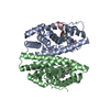
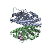
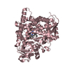
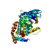
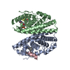
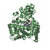
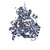
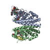
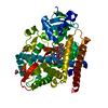
| Title | Distinct Monooxygenase and Farnesene Synthase Active Sites in Cytochrome P450 170A1 | ||||||
 Components Components | Putative cytochrome P450 | ||||||
 Keywords Keywords | OXIDOREDUCTASE / streptomyces / Cytochrome P450 oxidoreductase / CYP170A1 / antibiotic biosynthesis / farnesene synthase / Heme / Iron / Metal-binding / Monooxygenase | ||||||
| Function / homology |  Function and homology informationepi-isozizaene 5-monooxygenase / beta-farnesene synthase / sesquiterpene synthase activity / sesquiterpene biosynthetic process / ketone biosynthetic process / terpene synthase activity / oxidoreductase activity, acting on paired donors, with incorporation or reduction of molecular oxygen / antibiotic biosynthetic process / monooxygenase activity / iron ion binding ...epi-isozizaene 5-monooxygenase / beta-farnesene synthase / sesquiterpene synthase activity / sesquiterpene biosynthetic process / ketone biosynthetic process / terpene synthase activity / oxidoreductase activity, acting on paired donors, with incorporation or reduction of molecular oxygen / antibiotic biosynthetic process / monooxygenase activity / iron ion binding / heme binding / cytosol Function and homology informationepi-isozizaene 5-monooxygenase / beta-farnesene synthase / sesquiterpene synthase activity / sesquiterpene biosynthetic process / ketone biosynthetic process / terpene synthase activity / oxidoreductase activity, acting on paired donors, with incorporation or reduction of molecular oxygen / antibiotic biosynthetic process / monooxygenase activity / iron ion binding ...epi-isozizaene 5-monooxygenase / beta-farnesene synthase / sesquiterpene synthase activity / sesquiterpene biosynthetic process / ketone biosynthetic process / terpene synthase activity / oxidoreductase activity, acting on paired donors, with incorporation or reduction of molecular oxygen / antibiotic biosynthetic process / monooxygenase activity / iron ion binding / heme binding / cytosolSimilarity search - Function | ||||||
| Biological species |  Streptomyces coelicolor (bacteria) Streptomyces coelicolor (bacteria) | ||||||
| Method | X-RAY DIFFRACTION / SYNCHROTRON / MOLECULAR REPLACEMENT / Resolution: 3.3 Å | ||||||
 Authors Authors | Zhao, B. / Waterman, M.R. | ||||||
 Citation Citation | Journal: J.Biol.Chem. / Year: 2009 Title: Crystal structure of albaflavenone monooxygenase containing a moonlighting terpene synthase active site Authors: Zhao, B. / Lei, L. / Vassylyev, D.G. / Lin, X. / Cane, D.E. / Kelly, S.L. / Yuan, H. / Lamb, D.C. / Waterman, M.R. | ||||||
| History |
|
- Structure visualization
| Structure viewer | Molecule: MolmilJmol/JSmol |
|---|
- Downloads & links
-Download
| PDBx/mmCIF format | 3el3.cif.gz | 175.3 KB | Display | PDBx/mmCIF format |
|---|---|---|---|---|
| PDB format | pdb3el3.ent.gz | 138.5 KB | Display | PDB format |
| PDBx/mmJSON format | 3el3.json.gz | Tree view | PDBx/mmJSON format | |
| Others |  Other downloads Other downloads |
-Validation report
| Arichive directory | https://data.pdbj.org/pub/pdb/validation_reports/el/3el3ftp://data.pdbj.org/pub/pdb/validation_reports/el/3el3 | HTTPS FTP |
|---|
-Related structure data
| Related structure data |  3dbgSC S: Starting model for refinement C: citing same article ( |
|---|---|
| Similar structure data |

-Links
 PDBj
PDBj
- Assembly
| Deposited unit | 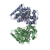
| ||||||||
|---|---|---|---|---|---|---|---|---|---|
| 1 | 
| ||||||||
| 2 | 
| ||||||||
| Unit cell |
|
-Components
| #1: Protein | Mass: 51837.141 Da / Num. of mol.: 2 Source method: isolated from a genetically manipulated source Source: (gene. exp.) Streptomyces coelicolor (bacteria) / Plasmid: pET17b / Production host: Escherichia coli (E. coli) / Strain (production host): BL21(DE3) / References: UniProt: Q9K498#2: Chemical | Heme B  Mass: 616.487 Da / Num. of mol.: 2 / Source method: obtained synthetically / Formula: C34H32FeN4O4 Mass: 616.487 Da / Num. of mol.: 2 / Source method: obtained synthetically / Formula: C34H32FeN4O4#3: Chemical | ChemComp-EL3 / (   Mass: 204.351 Da / Num. of mol.: 4 / Source method: obtained synthetically / Formula: C15H24 Mass: 204.351 Da / Num. of mol.: 4 / Source method: obtained synthetically / Formula: C15H24 |
|---|
-Experimental details
-Experiment
| Experiment | Method: X-RAY DIFFRACTION / Number of used crystals: 8 |
|---|
- Sample preparation
| Crystal | Density Matthews: 2.28 Å3/Da / Density % sol: 46.08 % |
|---|---|
| Crystal grow | Temperature: 295 K / Method: vapor diffusion, hanging drop / pH: 7 Details: 2.0 M ammonium sulphate, 0.1M Tris-HCl pH7.0, 1mM epi-isozizaene, VAPOR DIFFUSION, HANGING DROP, temperature 295K |
-Data collection
| Diffraction | Mean temperature: 200 K |
|---|---|
| Diffraction source | Source: SYNCHROTRON / Site: APS  / Beamline: 21-ID-G / Wavelength: 1 Å / Beamline: 21-ID-G / Wavelength: 1 Å |
| Detector | Type: MARMOSAIC 300 mm CCD / Detector: CCD / Date: Jun 19, 2008 |
| Radiation | Monochromator: Si 220 CHANNEL / Protocol: SINGLE WAVELENGTH / Monochromatic (M) / Laue (L): M / Scattering type: x-ray |
| Radiation wavelength | Wavelength: 1 Å / Relative weight: 1 |
| Reflection | Resolution: 3.3→50 Å / Num. obs: 12957 / % possible obs: 80.1 % / Observed criterion σ(F): 2 / Observed criterion σ(I): 2 / Redundancy: 3.7 % / Rmerge(I) obs: 0.064 / Rsym value: 0.039 |
| Reflection shell | Resolution: 3.3→3.41 Å / Redundancy: 2.1 % / Rmerge(I) obs: 0.244 / Mean I/σ(I) obs: 2.37 / Num. unique all: 1024 / Rsym value: 0.259 / % possible all: 64.3 |
- Processing
| Software |
| |||||||||||||||||||||||||
|---|---|---|---|---|---|---|---|---|---|---|---|---|---|---|---|---|---|---|---|---|---|---|---|---|---|---|
| Refinement | Method to determine structure: MOLECULAR REPLACEMENT Starting model: PDB entry 3DBG Resolution: 3.3→35.48 Å / Isotropic thermal model: Isotropic / Cross valid method: THROUGHOUT / σ(F): 0.2 / σ(I): 0.2 / Stereochemistry target values: Engh & Huber
| |||||||||||||||||||||||||
| Displacement parameters | Biso mean: 88.52 Å2
| |||||||||||||||||||||||||
| Refine analyze |
| |||||||||||||||||||||||||
| Refinement step | Cycle: LAST / Resolution: 3.3→35.48 Å
| |||||||||||||||||||||||||
| Refine LS restraints |
| |||||||||||||||||||||||||
| LS refinement shell | Resolution: 3.3→3.45 Å / Rfactor Rfree error: 0.01
|
