 Movie
Movie Controller

[English] 日本語
 Yorodumi
- PDB-3e82: Crystal structure of a putative oxidoreductase from Klebsiella pn... -
+ Open data

- Basic information
| Entry | Database: PDB / ID: 3.0E+82 | ||||||
|---|---|---|---|---|---|---|---|
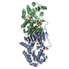
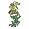
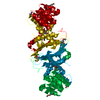
| Title | Crystal structure of a putative oxidoreductase from Klebsiella pneumoniae | ||||||
 Components Components | Putative oxidoreductase | ||||||
 Keywords Keywords | OXIDOREDUCTASE / NAD / GFO/IDH/MOCA family / PSI-2 / NYSGXRC / 11136f / Structural Genomics / Protein Structure Initiative / New York SGX Research Center for Structural Genomics | ||||||
| Function / homology |  Function and homology information Function and homology information | ||||||
| Biological species |  Klebsiella pneumoniae subsp. pneumoniae MGH 78578 (bacteria) Klebsiella pneumoniae subsp. pneumoniae MGH 78578 (bacteria) | ||||||
| Method | X-RAY DIFFRACTION / SYNCHROTRON / SAD / Resolution: 2.04 Å | ||||||
 Authors Authors | Eswaramoorthy, S. / Mohammad, M.B. / Thomas, C.A. / Brown, A.C. / Burley, S.K. / Swaminathan, S. / New York SGX Research Center for Structural Genomics (NYSGXRC) | ||||||
 Citation Citation | Journal: To be Published Title: Crystal structure of a putative oxidoreductase from Klebsiella pneumoniae Authors: Eswaramoorthy, S. / Mohammad, M.B. / Thomas, C.A. / Brown, A.C. / Burley, S.K. / Swaminathan, S. | ||||||
| History |
|
- Structure visualization
| Structure viewer | Molecule: MolmilJmol/JSmol |
|---|
- Downloads & links
-Download
| PDBx/mmCIF format | 3e82.cif.gz | 279.2 KB | Display | PDBx/mmCIF format |
|---|---|---|---|---|
| PDB format | pdb3e82.ent.gz | 234.2 KB | Display | PDB format |
| PDBx/mmJSON format | 3e82.json.gz | Tree view | PDBx/mmJSON format | |
| Others |  Other downloads Other downloads |
-Validation report
| Arichive directory | https://data.pdbj.org/pub/pdb/validation_reports/e8/3e82ftp://data.pdbj.org/pub/pdb/validation_reports/e8/3e82 | HTTPS FTP |
|---|
-Related structure data
| Similar structure data | |
|---|---|
| Other databases |


-Links
 PDBj
PDBj- Assembly
| Deposited unit | 
| ||||||||
|---|---|---|---|---|---|---|---|---|---|
| 1 | 
| ||||||||
| 2 | 
| ||||||||
| Unit cell |
|
-Components
| #1: Protein | Mass: 40390.641 Da / Num. of mol.: 4 Source method: isolated from a genetically manipulated source Source: (gene. exp.) Klebsiella pneumoniae subsp. pneumoniae MGH 78578 (bacteria)Gene: KPN78578_13420, KPN_01371 / Plasmid: BC-PSGX3(BC), TOP10-INVITROGEN / Production host: Escherichia coli (E. coli) / Strain (production host): BL21 (DE3)-CODON+RIL-STRATAGENE / References: UniProt: A6T882#2: Chemical | ChemComp-CL / Chloride  Mass: 35.453 Da / Num. of mol.: 4 / Source method: obtained synthetically / Formula: Cl Mass: 35.453 Da / Num. of mol.: 4 / Source method: obtained synthetically / Formula: Cl#3: Water | ChemComp-HOH / | Water Mass: 18.015 Da / Num. of mol.: 684 / Source method: isolated from a natural source / Formula: H2O Mass: 18.015 Da / Num. of mol.: 684 / Source method: isolated from a natural source / Formula: H2O |
|---|
-Experimental details
-Experiment
| Experiment | Method: X-RAY DIFFRACTION / Number of used crystals: 1 |
|---|
- Sample preparation
| Crystal | Density Matthews: 2.72 Å3/Da / Density % sol: 54.85 % |
|---|---|
| Crystal grow | Temperature: 291 K / Method: vapor diffusion, sitting drop / pH: 5.5 Details: 25% PEG3350, 0.1M Bis-Tris, pH 5.5, VAPOR DIFFUSION, SITTING DROP, temperature 291K |
-Data collection
| Diffraction | Mean temperature: 100 K |
|---|---|
| Diffraction source | Source: SYNCHROTRON / Site: NSLS  / Beamline: X12C / Wavelength: 0.979 Å / Beamline: X12C / Wavelength: 0.979 Å |
| Detector | Type: ADSC QUANTUM 210 / Detector: CCD / Date: Jun 30, 2008 / Details: mirrors |
| Radiation | Monochromator: Si 111 CHANNEL / Protocol: SINGLE WAVELENGTH / Monochromatic (M) / Laue (L): M / Scattering type: x-ray |
| Radiation wavelength | Wavelength: 0.979 Å / Relative weight: 1 |
| Reflection | Resolution: 2.04→50 Å / Num. all: 109371 / Num. obs: 109371 / % possible obs: 98.8 % / Observed criterion σ(F): 0 / Observed criterion σ(I): 0 / Redundancy: 7.5 % / Biso Wilson estimate: 16.4 Å2 / Rsym value: 0.096 / Net I/σ(I): 13 |
| Reflection shell | Resolution: 2.04→2.11 Å / Redundancy: 6.6 % / Rmerge(I) obs: 0.56 / Num. unique all: 10624 / % possible all: 97.1 |
- Processing
| Software |
| |||||||||||||||||||||||||
|---|---|---|---|---|---|---|---|---|---|---|---|---|---|---|---|---|---|---|---|---|---|---|---|---|---|---|
| Refinement | Method to determine structure: SAD / Resolution: 2.04→35.87 Å / Isotropic thermal model: Isotropic / Cross valid method: THROUGHOUT / σ(F): 0 / σ(I): 0 / Stereochemistry target values: Engh & Huber
| |||||||||||||||||||||||||
| Displacement parameters | Biso mean: 28.9 Å2
| |||||||||||||||||||||||||
| Refine analyze |
| |||||||||||||||||||||||||
| Refinement step | Cycle: LAST / Resolution: 2.04→35.87 Å
| |||||||||||||||||||||||||
| Refine LS restraints |
| |||||||||||||||||||||||||
| LS refinement shell | Resolution: 2.04→2.17 Å / Rfactor Rfree error: 0.012
|
