 Movie
Movie Controller

[English] 日本語
 Yorodumi
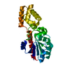
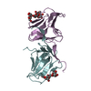
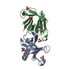
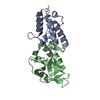
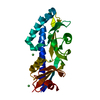
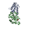
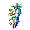
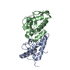
- PDB-3e46: Crystal structure of ubiquitin-conjugating enzyme E2-25kDa (Hunti... -
+ Open data

- Basic information
| Entry | Database: PDB / ID: 3.0E+46 | ||||||
|---|---|---|---|---|---|---|---|
| Title | Crystal structure of ubiquitin-conjugating enzyme E2-25kDa (Huntington interacting protein 2) M172A mutant | ||||||
 Components Components | Ubiquitin-conjugating enzyme E2-25 kDa | ||||||
 Keywords Keywords |  LIGASE / Ubiquitin-Conjugating / Huntington Interacting / E2-25K / Alternative splicing / Cytoplasm / Ubl conjugation / Ubl conjugation pathway LIGASE / Ubiquitin-Conjugating / Huntington Interacting / E2-25K / Alternative splicing / Cytoplasm / Ubl conjugation / Ubl conjugation pathway | ||||||
| Function / homology |  Function and homology information Function and homology informationfree ubiquitin chain polymerization / regulation of proteasomal ubiquitin-dependent protein catabolic process / filopodium tip / positive regulation of tumor necrosis factor-mediated signaling pathway / positive regulation of type I interferon-mediated signaling pathway / ubiquitin-ubiquitin ligase activity / E2 ubiquitin-conjugating enzyme / ubiquitin conjugating enzyme activity / cellular response to interferon-beta / protein K48-linked ubiquitination ...free ubiquitin chain polymerization / regulation of proteasomal ubiquitin-dependent protein catabolic process / filopodium tip / positive regulation of tumor necrosis factor-mediated signaling pathway / positive regulation of type I interferon-mediated signaling pathway / ubiquitin-ubiquitin ligase activity / E2 ubiquitin-conjugating enzyme / ubiquitin conjugating enzyme activity / cellular response to interferon-beta / protein K48-linked ubiquitination / Synthesis of active ubiquitin: roles of E1 and E2 enzymes / positive regulation of peptidyl-threonine phosphorylation / Negative regulators of DDX58/IFIH1 signaling / protein polyubiquitination / ubiquitin-protein transferase activity / Antigen processing: Ubiquitination & Proteasome degradation / ubiquitin-dependent protein catabolic process / proteasome-mediated ubiquitin-dependent protein catabolic process / ubiquitin protein ligase binding / ATP binding / nucleus / cytosol / cytoplasmSimilarity search - Function | ||||||
| Biological species |  Homo sapiens (human) Homo sapiens (human) | ||||||
| Method | X-RAY DIFFRACTION / SYNCHROTRON / MOLECULAR REPLACEMENT / molecular replacement / Resolution: 1.86 Å | ||||||
 Authors Authors | Hughes, R.C. / Wilson, R.C. / Flatt, J.W. / Meehan, E.J. / Ng, J.D. / Twigg, P.D. | ||||||
 Citation Citation | Journal: Acta Crystallogr.,Sect.F / Year: 2009 Title: Structure of full-length ubiquitin-conjugating enzyme E2-25K (huntingtin-interacting protein 2). Authors: Wilson, R.C. / Hughes, R.C. / Flatt, J.W. / Meehan, E.J. / Ng, J.D. / Twigg, P.D. | ||||||
| History |
|
- Structure visualization
| Structure viewer | Molecule: MolmilJmol/JSmol |
|---|
- Downloads & links
-Download
| PDBx/mmCIF format | 3e46.cif.gz | 61.7 KB | Display | PDBx/mmCIF format |
|---|---|---|---|---|
| PDB format | pdb3e46.ent.gz | 42.2 KB | Display | PDB format |
| PDBx/mmJSON format | 3e46.json.gz | Tree view | PDBx/mmJSON format | |
| Others |  Other downloads Other downloads |
-Validation report
| Arichive directory | https://data.pdbj.org/pub/pdb/validation_reports/e4/3e46ftp://data.pdbj.org/pub/pdb/validation_reports/e4/3e46 | HTTPS FTP |
|---|
-Related structure data
| Related structure data | 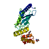 3f92C  2bepS S: Starting model for refinement C: citing same article ( |
|---|---|
| Similar structure data |


-Links
 PDBj
PDBj


- Assembly
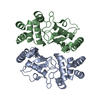
| Deposited unit | 
| ||||||||
|---|---|---|---|---|---|---|---|---|---|
| 1 |
| ||||||||
| Unit cell |
|
-Components
| #1: Protein | Mass: 28205.840 Da / Num. of mol.: 1 / Mutation: M172A Source method: isolated from a genetically manipulated source Source: (gene. exp.) Homo sapiens (human) / Gene: UBE2K, HIP2, LIG / Plasmid: pET30a / Production host:  Escherichia coli (E. coli) / Strain (production host): BL21(DE3) / References: UniProt: P61086, ubiquitin-protein ligase Escherichia coli (E. coli) / Strain (production host): BL21(DE3) / References: UniProt: P61086, ubiquitin-protein ligase |
|---|---|
| #2: Chemical | ChemComp-CA /   Mass: 40.078 Da / Num. of mol.: 1 / Source method: obtained synthetically / Formula: Ca Mass: 40.078 Da / Num. of mol.: 1 / Source method: obtained synthetically / Formula: Ca |
| #3: Water | ChemComp-HOH / Water Mass: 18.015 Da / Num. of mol.: 207 / Source method: isolated from a natural source / Formula: H2O Mass: 18.015 Da / Num. of mol.: 207 / Source method: isolated from a natural source / Formula: H2O |
-Experimental details
-Experiment
| Experiment | Method: X-RAY DIFFRACTION / Number of used crystals: 1 |
|---|
- Sample preparation
| Crystal | Density Matthews: 3.08 Å3/Da / Density % sol: 60.04 % |
|---|---|
| Crystal grow | Temperature: 298 K / Method: vapor diffusion, sitting drop / pH: 6 Details: Calcium acetate, Sodium acetate, NaCl, PEG 8000, pH 6.0, VAPOR DIFFUSION, SITTING DROP, temperature 298K |
-Data collection
| Diffraction | Mean temperature: 100 K | |||||||||||||||||||||||||||||||||||||||||||||||||||||||||||||||||||||||||||||
|---|---|---|---|---|---|---|---|---|---|---|---|---|---|---|---|---|---|---|---|---|---|---|---|---|---|---|---|---|---|---|---|---|---|---|---|---|---|---|---|---|---|---|---|---|---|---|---|---|---|---|---|---|---|---|---|---|---|---|---|---|---|---|---|---|---|---|---|---|---|---|---|---|---|---|---|---|---|---|
| Diffraction source | Source: SYNCHROTRON / Site: APS  / Beamline: 22-ID / Wavelength: 0.97 Å / Beamline: 22-ID / Wavelength: 0.97 Å | |||||||||||||||||||||||||||||||||||||||||||||||||||||||||||||||||||||||||||||
| Detector | Type: MARMOSAIC 300 mm CCD / Detector: CCD / Date: Jul 7, 2008 | |||||||||||||||||||||||||||||||||||||||||||||||||||||||||||||||||||||||||||||
| Radiation | Protocol: SINGLE WAVELENGTH / Monochromatic (M) / Laue (L): M / Scattering type: x-ray | |||||||||||||||||||||||||||||||||||||||||||||||||||||||||||||||||||||||||||||
| Radiation wavelength | Wavelength: 0.97 Å / Relative weight: 1 | |||||||||||||||||||||||||||||||||||||||||||||||||||||||||||||||||||||||||||||
| Reflection | Redundancy: 4 % / Number: 109190 / Rmerge(I) obs: 0.081 / Χ2: 1.35 / D res high: 1.84 Å / D res low: 50 Å / Num. obs: 27414 / % possible obs: 90.4 | |||||||||||||||||||||||||||||||||||||||||||||||||||||||||||||||||||||||||||||
| Diffraction reflection shell |
| |||||||||||||||||||||||||||||||||||||||||||||||||||||||||||||||||||||||||||||
| Reflection | Resolution: 1.86→50 Å / Num. obs: 27414 / % possible obs: 93.4 % / Observed criterion σ(I): 2 / Redundancy: 4 % / Biso Wilson estimate: 26 Å2 / Rsym value: 0.081 | |||||||||||||||||||||||||||||||||||||||||||||||||||||||||||||||||||||||||||||
| Reflection shell | Resolution: 1.86→1.91 Å / Redundancy: 1.4 % / Mean I/σ(I) obs: 1.98 / Num. unique all: 1674 / Rsym value: 0.374 / % possible all: 52.4 |
-Phasing
| Phasing | Method: molecular replacement | |||||||||
|---|---|---|---|---|---|---|---|---|---|---|
| Phasing MR |
|
- Processing
| Software |
| ||||||||||||||||||||||||||||||||||||||||||||||||||||||||||||||||||||||||||||||||||||||||||||||||||||||||||||||||||||||||||||||||||
|---|---|---|---|---|---|---|---|---|---|---|---|---|---|---|---|---|---|---|---|---|---|---|---|---|---|---|---|---|---|---|---|---|---|---|---|---|---|---|---|---|---|---|---|---|---|---|---|---|---|---|---|---|---|---|---|---|---|---|---|---|---|---|---|---|---|---|---|---|---|---|---|---|---|---|---|---|---|---|---|---|---|---|---|---|---|---|---|---|---|---|---|---|---|---|---|---|---|---|---|---|---|---|---|---|---|---|---|---|---|---|---|---|---|---|---|---|---|---|---|---|---|---|---|---|---|---|---|---|---|---|---|
| Refinement | Method to determine structure: MOLECULAR REPLACEMENT Starting model: PDB entry 2BEP Resolution: 1.86→32.38 Å / Cor.coef. Fo:Fc: 0.968 / Cor.coef. Fo:Fc free: 0.953 / WRfactor Rfree: 0.22 / WRfactor Rwork: 0.171 / Occupancy max: 1 / Occupancy min: 0 / FOM work R set: 0.846 / SU B: 2.829 / SU ML: 0.081 / SU R Cruickshank DPI: 0.103 / SU Rfree: 0.107 / Cross valid method: THROUGHOUT / σ(F): 0 / ESU R: 0.103 / ESU R Free: 0.107 / Stereochemistry target values: MAXIMUM LIKELIHOOD
| ||||||||||||||||||||||||||||||||||||||||||||||||||||||||||||||||||||||||||||||||||||||||||||||||||||||||||||||||||||||||||||||||||
| Solvent computation | Ion probe radii: 0.8 Å / Shrinkage radii: 0.8 Å / VDW probe radii: 1.2 Å / Solvent model: BABINET MODEL WITH MASK | ||||||||||||||||||||||||||||||||||||||||||||||||||||||||||||||||||||||||||||||||||||||||||||||||||||||||||||||||||||||||||||||||||
| Displacement parameters | Biso max: 269.48 Å2 / Biso mean: 36.498 Å2 / Biso min: 17.68 Å2
| ||||||||||||||||||||||||||||||||||||||||||||||||||||||||||||||||||||||||||||||||||||||||||||||||||||||||||||||||||||||||||||||||||
| Refinement step | Cycle: LAST / Resolution: 1.86→32.38 Å
| ||||||||||||||||||||||||||||||||||||||||||||||||||||||||||||||||||||||||||||||||||||||||||||||||||||||||||||||||||||||||||||||||||
| Refine LS restraints |
| ||||||||||||||||||||||||||||||||||||||||||||||||||||||||||||||||||||||||||||||||||||||||||||||||||||||||||||||||||||||||||||||||||
| LS refinement shell | Resolution: 1.86→1.91 Å / Total num. of bins used: 20
|
