 Movie
Movie Controller

[English] 日本語
 Yorodumi
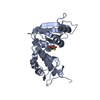
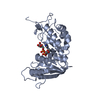
- PDB-3dzf: Crystal structure of human CD38 extracellular domain complexed wi... -
+ Open data

- Basic information
| Entry | Database: PDB / ID: 3dzf | ||||||
|---|---|---|---|---|---|---|---|
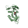
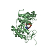
| Title | Crystal structure of human CD38 extracellular domain complexed with a covalent intermediate, ara-F-ribose-5'-phosphate | ||||||
 Components Components | ADP-ribosyl cyclase 1 Cyclic ADP-ribose Cyclic ADP-ribose | ||||||
 Keywords Keywords | HYDROLASE / covalent intermediate / BETA SHEETS / ALPHA BUNDLE / Diabetes mellitus / Glycoprotein / Membrane / NAD / Receptor / Signal-anchor / Transmembrane | ||||||
| Function / homology |  Function and homology information Function and homology information2'-phospho-ADP-ribosyl cyclase/2'-phospho-cyclic-ADP-ribose transferase / phosphorus-oxygen lyase activity / artery smooth muscle contraction / Nicotinate metabolism / NAD+ nucleosidase activity / ADP-ribosyl cyclase/cyclic ADP-ribose hydrolase / NAD metabolic process / NAD+ nucleotidase, cyclic ADP-ribose generating / negative regulation of bone resorption / response to hydroperoxide ...2'-phospho-ADP-ribosyl cyclase/2'-phospho-cyclic-ADP-ribose transferase / phosphorus-oxygen lyase activity / artery smooth muscle contraction / Nicotinate metabolism / NAD+ nucleosidase activity / ADP-ribosyl cyclase/cyclic ADP-ribose hydrolase / NAD metabolic process / NAD+ nucleotidase, cyclic ADP-ribose generating / negative regulation of bone resorption / response to hydroperoxide / long-term synaptic depression / Hydrolases; Glycosylases; Hydrolysing N-glycosyl compounds / B cell proliferation / response to retinoic acid / positive regulation of B cell proliferation / positive regulation of vasoconstriction / response to interleukin-1 / response to progesterone / female pregnancy / apoptotic signaling pathway / B cell receptor signaling pathway / positive regulation of insulin secretion / response to estradiol / negative regulation of neuron projection development / positive regulation of cytosolic calcium ion concentration / transferase activity / positive regulation of cell growth / basolateral plasma membrane / nuclear membrane / response to hypoxia / response to xenobiotic stimulus / negative regulation of DNA-templated transcription / negative regulation of apoptotic process / positive regulation of DNA-templated transcription / cell surface / signal transduction / extracellular exosome / membrane / identical protein binding / plasma membraneSimilarity search - Function | ||||||
| Biological species |  Homo sapiens (human) Homo sapiens (human) | ||||||
| Method | X-RAY DIFFRACTION / SYNCHROTRON / MOLECULAR REPLACEMENT / Resolution: 2.01 Å | ||||||
 Authors Authors | Liu, Q. / Kriksunov, I.A. / Jiang, H. / Graeff, R. / Lin, H. / Lee, H.C. / Hao, Q. | ||||||
 Citation Citation | Journal: Chem.Biol. / Year: 2008 Title: Covalent and Noncovalent Intermediates of an NAD Utilizing Enzyme, Human CD38. Authors: Liu, Q. / Kriksunov, I.A. / Jiang, H. / Graeff, R. / Lin, H. / Lee, H.C. / Hao, Q. | ||||||
| History |
|
- Structure visualization
| Structure viewer | Molecule: MolmilJmol/JSmol |
|---|
- Downloads & links
-Download
| PDBx/mmCIF format | 3dzf.cif.gz | 322.2 KB | Display | PDBx/mmCIF format |
|---|---|---|---|---|
| PDB format | pdb3dzf.ent.gz | 262.4 KB | Display | PDB format |
| PDBx/mmJSON format | 3dzf.json.gz | Tree view | PDBx/mmJSON format | |
| Others |  Other downloads Other downloads |
-Validation report
| Arichive directory | https://data.pdbj.org/pub/pdb/validation_reports/dz/3dzfftp://data.pdbj.org/pub/pdb/validation_reports/dz/3dzf | HTTPS FTP |
|---|
-Related structure data
| Related structure data |  3dzgC  3dzhC  3dziC  3dzjC  3dzkC  1yh3S C: citing same article ( S: Starting model for refinement |
|---|---|
| Similar structure data |


-Links
 PDBj
PDBj
- Assembly
| Deposited unit | 
| ||||||||
|---|---|---|---|---|---|---|---|---|---|
| 1 | 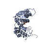
| ||||||||
| 2 | 
| ||||||||
| 3 | 
| ||||||||
| 4 | 
| ||||||||
| 5 | 
| ||||||||
| 6 | 
| ||||||||
| Unit cell |
|
-Components
| #1: Protein | Cyclic ADP-ribose / Cyclic ADP-ribose hydrolase 1 / cADPr hydrolase 1 / T10 / CD38 antigen Mass: 30380.393 Da / Num. of mol.: 6 / Fragment: Enzymatic domain: UNP residues 45-300 / Mutation: Q49T, N100D, N164D, N209D, N219D Source method: isolated from a genetically manipulated source Source: (gene. exp.) Homo sapiens (human) / Gene: CD38 / Plasmid: pPICZ(alpha)A / Production host:  Pichia pastoris (fungus) / Strain (production host): X-33 (Invitrogen) / References: UniProt: P28907, NAD+ glycohydrolase Pichia pastoris (fungus) / Strain (production host): X-33 (Invitrogen) / References: UniProt: P28907, NAD+ glycohydrolase#2: Sugar | ChemComp-RF5 /   Type: D-saccharide, alpha linking / Mass: 232.101 Da / Num. of mol.: 6 / Source method: obtained synthetically / Formula: C5H10FO7P Type: D-saccharide, alpha linking / Mass: 232.101 Da / Num. of mol.: 6 / Source method: obtained synthetically / Formula: C5H10FO7P#3: Water | ChemComp-HOH / | Water Mass: 18.015 Da / Num. of mol.: 609 / Source method: isolated from a natural source / Formula: H2O Mass: 18.015 Da / Num. of mol.: 609 / Source method: isolated from a natural source / Formula: H2ONonpolymer details | AUTHORS STATE THAT THE LIGAND RF5 IN THIS ENTRY IS A REACTION INTERMEDIATE, WITH THE (C1') ATOM ...AUTHORS STATE THAT THE LIGAND RF5 IN THIS ENTRY IS A REACTION INTERMEDIA | |
|---|
-Experimental details
-Experiment
| Experiment | Method: X-RAY DIFFRACTION / Number of used crystals: 1 |
|---|
- Sample preparation
| Crystal | Density Matthews: 2.22 Å3/Da / Density % sol: 44.69 % |
|---|---|
| Crystal grow | Temperature: 298 K / pH: 6 Details: 100 mM MES pH 6.0, 15% PEG 4000, VAPOR DIFFUSION, HANGING DROP, temperature 298.0K |
-Data collection
| Diffraction | Mean temperature: 100 K |
|---|---|
| Diffraction source | Source: SYNCHROTRON / Site: CHESS  / Beamline: A1 / Wavelength: 0.9778 / Beamline: A1 / Wavelength: 0.9778 |
| Detector | Type: ADSC QUANTUM 210 / Detector: CCD / Date: Apr 1, 2007 |
| Radiation | Monochromator: SI 111 CHANNEL / Protocol: SINGLE WAVELENGTH / Monochromatic (M) / Laue (L): M / Scattering type: x-ray |
| Radiation wavelength | Wavelength: 0.9778 Å / Relative weight: 1 |
| Reflection | Resolution: 2→30 Å / Num. obs: 90177 / % possible obs: 86.7 % / Observed criterion σ(I): 2 / Redundancy: 3.6 % / Rmerge(I) obs: 0.064 / Rsym value: 0.064 / Net I/σ(I): 18.1 |
| Reflection shell | Resolution: 2→2.07 Å / Redundancy: 3.2 % / Rmerge(I) obs: 0.485 / Mean I/σ(I) obs: 2.5 / Rsym value: 0.485 / % possible all: 75 |
- Processing
| Software |
| |||||||||||||||||||||||||||||||||||||||||||||||||||||||||||||||||||||||||||||||||||||||||||||||||||||||||||||||||||||||||||||||||||||||||||||||||||||||||||||||||||||||||||||||
|---|---|---|---|---|---|---|---|---|---|---|---|---|---|---|---|---|---|---|---|---|---|---|---|---|---|---|---|---|---|---|---|---|---|---|---|---|---|---|---|---|---|---|---|---|---|---|---|---|---|---|---|---|---|---|---|---|---|---|---|---|---|---|---|---|---|---|---|---|---|---|---|---|---|---|---|---|---|---|---|---|---|---|---|---|---|---|---|---|---|---|---|---|---|---|---|---|---|---|---|---|---|---|---|---|---|---|---|---|---|---|---|---|---|---|---|---|---|---|---|---|---|---|---|---|---|---|---|---|---|---|---|---|---|---|---|---|---|---|---|---|---|---|---|---|---|---|---|---|---|---|---|---|---|---|---|---|---|---|---|---|---|---|---|---|---|---|---|---|---|---|---|---|---|---|---|---|
| Refinement | Method to determine structure: MOLECULAR REPLACEMENT Starting model: PDB ENTRY 1YH3 Resolution: 2.01→20 Å / Cor.coef. Fo:Fc: 0.962 / Cor.coef. Fo:Fc free: 0.928 / SU B: 10.472 / SU ML: 0.149 / TLS residual ADP flag: LIKELY RESIDUAL / Cross valid method: THROUGHOUT / σ(F): 0 / ESU R: 0.269 / ESU R Free: 0.228 / Stereochemistry target values: MAXIMUM LIKELIHOOD
| |||||||||||||||||||||||||||||||||||||||||||||||||||||||||||||||||||||||||||||||||||||||||||||||||||||||||||||||||||||||||||||||||||||||||||||||||||||||||||||||||||||||||||||||
| Solvent computation | Ion probe radii: 0.8 Å / Shrinkage radii: 0.8 Å / VDW probe radii: 1.2 Å / Solvent model: BABINET MODEL WITH MASK | |||||||||||||||||||||||||||||||||||||||||||||||||||||||||||||||||||||||||||||||||||||||||||||||||||||||||||||||||||||||||||||||||||||||||||||||||||||||||||||||||||||||||||||||
| Displacement parameters | Biso mean: 38.75 Å2
| |||||||||||||||||||||||||||||||||||||||||||||||||||||||||||||||||||||||||||||||||||||||||||||||||||||||||||||||||||||||||||||||||||||||||||||||||||||||||||||||||||||||||||||||
| Refinement step | Cycle: LAST / Resolution: 2.01→20 Å
| |||||||||||||||||||||||||||||||||||||||||||||||||||||||||||||||||||||||||||||||||||||||||||||||||||||||||||||||||||||||||||||||||||||||||||||||||||||||||||||||||||||||||||||||
| Refine LS restraints |
| |||||||||||||||||||||||||||||||||||||||||||||||||||||||||||||||||||||||||||||||||||||||||||||||||||||||||||||||||||||||||||||||||||||||||||||||||||||||||||||||||||||||||||||||
| LS refinement shell | Resolution: 2.01→2.06 Å / Total num. of bins used: 20
| |||||||||||||||||||||||||||||||||||||||||||||||||||||||||||||||||||||||||||||||||||||||||||||||||||||||||||||||||||||||||||||||||||||||||||||||||||||||||||||||||||||||||||||||
| Refinement TLS params. | Method: refined / Refine-ID: X-RAY DIFFRACTION
| |||||||||||||||||||||||||||||||||||||||||||||||||||||||||||||||||||||||||||||||||||||||||||||||||||||||||||||||||||||||||||||||||||||||||||||||||||||||||||||||||||||||||||||||
| Refinement TLS group |
|
