 Movie
Movie Controller

[English] 日本語
 Yorodumi
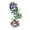
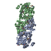
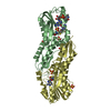
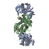
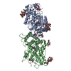
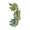
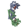
- PDB-3dzb: Crystal structure of Prephenate dehydrogenase from Streptococcus ... -
+ Open data

- Basic information
| Entry | Database: PDB / ID: 3dzb | ||||||
|---|---|---|---|---|---|---|---|
| Title | Crystal structure of Prephenate dehydrogenase from Streptococcus thermophilus | ||||||
 Components Components | Prephenate dehydrogenase | ||||||
 Keywords Keywords | BIOSYNTHETIC PROTEIN / domain swap / 11134a2 / PSI2 / NYSGXRC / tyrosine biosynthesis / Ec:1.3.12.- / Structural Genomics / Protein Structure Initiative / New York SGX Research Center for Structural Genomics / OXIDOREDUCTASE | ||||||
| Function / homology |  Function and homology informationprephenate dehydrogenase (NADP+) activity / prephenate dehydrogenase (NAD+) activity / tyrosine biosynthetic process Function and homology informationprephenate dehydrogenase (NADP+) activity / prephenate dehydrogenase (NAD+) activity / tyrosine biosynthetic processSimilarity search - Function | ||||||
| Biological species |  Streptococcus thermophilus LMG 18311 (bacteria) Streptococcus thermophilus LMG 18311 (bacteria) | ||||||
| Method | X-RAY DIFFRACTION / SYNCHROTRON / SAD / Resolution: 2.46 Å | ||||||
 Authors Authors | Zhang, Z. / Eswaramoorthy, S. / Burley, S.K. / Swaminathan, S. / New York SGX Research Center for Structural Genomics (NYSGXRC) | ||||||
 Citation Citation | Journal: To be Published Title: Crystal structure of Prephenate dehydrogenase from Streptococcus thermophilus Authors: Zhang, Z. / Eswaramoorthy, S. / Burley, S.K. / Swaminathan, S. | ||||||
| History |
|
- Structure visualization
| Structure viewer | Molecule: MolmilJmol/JSmol |
|---|
- Downloads & links
-Download
| PDBx/mmCIF format | 3dzb.cif.gz | 117.4 KB | Display | PDBx/mmCIF format |
|---|---|---|---|---|
| PDB format | pdb3dzb.ent.gz | 96.7 KB | Display | PDB format |
| PDBx/mmJSON format | 3dzb.json.gz | Tree view | PDBx/mmJSON format | |
| Others |  Other downloads Other downloads |
-Validation report
| Arichive directory | https://data.pdbj.org/pub/pdb/validation_reports/dz/3dzbftp://data.pdbj.org/pub/pdb/validation_reports/dz/3dzb | HTTPS FTP |
|---|
-Related structure data
| Similar structure data | |
|---|---|
| Other databases |


-Links
 PDBj
PDBj


- Assembly
| Deposited unit | 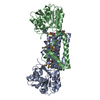
| ||||||||
|---|---|---|---|---|---|---|---|---|---|
| 1 |
| ||||||||
| Unit cell |
|
-Components
| #1: Protein | Mass: 35778.086 Da / Num. of mol.: 2 Source method: isolated from a genetically manipulated source Source: (gene. exp.) Streptococcus thermophilus LMG 18311 (bacteria)Gene: tyrA, stu0642 / Plasmid: BC-pSGX3(BC), TOP10-invitrogen / Production host: Escherichia coli (E. coli) / Strain (production host): BL21 (DE3)-Codon+RIL-stratagene / References: UniProt: Q5M554, prephenate dehydrogenase#2: Water | ChemComp-HOH / | Water Mass: 18.015 Da / Num. of mol.: 60 / Source method: isolated from a natural source / Formula: H2O Mass: 18.015 Da / Num. of mol.: 60 / Source method: isolated from a natural source / Formula: H2O |
|---|
-Experimental details
-Experiment
| Experiment | Method: X-RAY DIFFRACTION / Number of used crystals: 1 |
|---|
- Sample preparation
| Crystal | Density Matthews: 2.39 Å3/Da / Density % sol: 48.54 % |
|---|---|
| Crystal grow | Temperature: 291 K / Method: vapor diffusion, sitting drop / pH: 7.5 Details: 20% PEG 3350, 0.2 M Potassium Chloride, pH 7.5, VAPOR DIFFUSION, SITTING DROP, temperature 291K |
-Data collection
| Diffraction | Mean temperature: 100 K |
|---|---|
| Diffraction source | Source: SYNCHROTRON / Site: NSLS  / Beamline: X12C / Wavelength: 0.979 Å / Beamline: X12C / Wavelength: 0.979 Å |
| Detector | Type: ADSC QUANTUM 210 / Detector: CCD / Date: Jul 19, 2008 / Details: mirrors |
| Radiation | Monochromator: Si 111 CHANNEL / Protocol: SINGLE WAVELENGTH / Monochromatic (M) / Laue (L): M / Scattering type: x-ray |
| Radiation wavelength | Wavelength: 0.979 Å / Relative weight: 1 |
| Reflection | Resolution: 2.46→50 Å / Num. all: 25463 / Num. obs: 25463 / % possible obs: 99.9 % / Observed criterion σ(F): 0 / Observed criterion σ(I): 0 / Redundancy: 13.3 % / Biso Wilson estimate: 33.6 Å2 / Rmerge(I) obs: 0.086 / Net I/σ(I): 9.8 |
| Reflection shell | Resolution: 2.46→2.56 Å / Redundancy: 11.3 % / Rmerge(I) obs: 0.709 / Num. unique all: 2491 / % possible all: 99.4 |
- Processing
| Software |
| |||||||||||||||||||||||||
|---|---|---|---|---|---|---|---|---|---|---|---|---|---|---|---|---|---|---|---|---|---|---|---|---|---|---|
| Refinement | Method to determine structure: SAD / Resolution: 2.46→50 Å / Isotropic thermal model: isotropic / Cross valid method: THROUGHOUT / σ(F): 0 / σ(I): 0 / Stereochemistry target values: Engh & Huber
| |||||||||||||||||||||||||
| Displacement parameters | Biso mean: 38.9 Å2
| |||||||||||||||||||||||||
| Refine analyze |
| |||||||||||||||||||||||||
| Refinement step | Cycle: LAST / Resolution: 2.46→50 Å
| |||||||||||||||||||||||||
| Refine LS restraints |
| |||||||||||||||||||||||||
| LS refinement shell | Resolution: 2.46→2.57 Å / Rfactor Rfree error: 0.037
|
