 Movie
Movie Controller

[English] 日本語
 Yorodumi
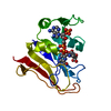
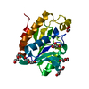
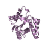
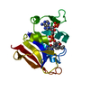
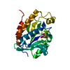
- PDB-3dwv: Glutathione peroxidase-type tryparedoxin peroxidase, oxidized form -
+ Open data

- Basic information
| Entry | Database: PDB / ID: 3dwv | ||||||
|---|---|---|---|---|---|---|---|
| Title | Glutathione peroxidase-type tryparedoxin peroxidase, oxidized form | ||||||
 Components Components | Glutathione peroxidase-like protein | ||||||
 Keywords Keywords |  OXIDOREDUCTASE / alpha beta / 3-Layer(aba) Sandwich / Glutaredoxin fold / Peroxidase OXIDOREDUCTASE / alpha beta / 3-Layer(aba) Sandwich / Glutaredoxin fold / Peroxidase | ||||||
| Function / homology |  Function and homology informationglycosome / glutathione peroxidase activity / response to oxidative stress Function and homology informationglycosome / glutathione peroxidase activity / response to oxidative stressSimilarity search - Function | ||||||
| Biological species |  Trypanosoma brucei (eukaryote) Trypanosoma brucei (eukaryote) | ||||||
| Method | X-RAY DIFFRACTION / SYNCHROTRON / MOLECULAR REPLACEMENT / Resolution: 1.41 Å | ||||||
 Authors Authors | Tews, I. / Sinning, I. / Krauth-Siegel, L. | ||||||
 Citation Citation | Journal: J.Biol.Chem. / Year: 2008 Title: Structural basis for a distinct catalytic mechanism in Trypanosoma brucei tryparedoxin peroxidase. Authors: Melchers, J. / Diechtierow, M. / Feher, K. / Sinning, I. / Tews, I. / Krauth-Siegel, R.L. / Muhle-Goll, C. | ||||||
| History |
|
- Structure visualization
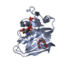
| Structure viewer | Molecule: MolmilJmol/JSmol |
|---|
- Downloads & links
-Download
| PDBx/mmCIF format | 3dwv.cif.gz | 166.6 KB | Display | PDBx/mmCIF format |
|---|---|---|---|---|
| PDB format | pdb3dwv.ent.gz | 129.4 KB | Display | PDB format |
| PDBx/mmJSON format | 3dwv.json.gz | Tree view | PDBx/mmJSON format | |
| Others |  Other downloads Other downloads |
-Validation report
| Arichive directory | https://data.pdbj.org/pub/pdb/validation_reports/dw/3dwvftp://data.pdbj.org/pub/pdb/validation_reports/dw/3dwv | HTTPS FTP |
|---|
-Related structure data
| Related structure data |  2rm5C 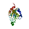 2rm6C  2gs3S C: citing same article ( S: Starting model for refinement |
|---|---|
| Similar structure data |


-Links
 PDBj
PDBj
- Assembly
| Deposited unit | 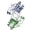
| ||||||||
|---|---|---|---|---|---|---|---|---|---|
| 1 | 
| ||||||||
| 2 | 
| ||||||||
| Unit cell |
|
-Components
| #1: Protein | Mass: 20983.975 Da / Num. of mol.: 2 / Mutation: C76S Source method: isolated from a genetically manipulated source Source: (gene. exp.) Trypanosoma brucei (eukaryote) / Gene: gpx3, Tb927.7.1140 / Plasmid: pQE30 / Production host:  Escherichia coli (E. coli) / Strain (production host): BL21(DE3) / References: UniProt: Q869A5, glutathione peroxidase Escherichia coli (E. coli) / Strain (production host): BL21(DE3) / References: UniProt: Q869A5, glutathione peroxidase#2: Water | ChemComp-HOH / | Water Mass: 18.015 Da / Num. of mol.: 360 / Source method: isolated from a natural source / Formula: H2O Mass: 18.015 Da / Num. of mol.: 360 / Source method: isolated from a natural source / Formula: H2OSequence details | THE VARIATIONS | |
|---|
-Experimental details
-Experiment
| Experiment | Method: X-RAY DIFFRACTION / Number of used crystals: 1 |
|---|
- Sample preparation
| Crystal | Density Matthews: 2.22 Å3/Da / Density % sol: 45.79 % / Mosaicity: 0.403 ° |
|---|---|
| Crystal grow | Temperature: 277 K / Method: vapor diffusion, hanging drop / pH: 4.6 Details: 2M ammonium sulphate, 0.1M sodium acetate, 1mM EDTA, pH 4.6, VAPOR DIFFUSION, HANGING DROP, temperature 277K |
-Data collection
| Diffraction | Mean temperature: 100 K |
|---|---|
| Diffraction source | Source: SYNCHROTRON / Site: ESRF  / Beamline: ID14-4 / Wavelength: 0.9393 Å / Beamline: ID14-4 / Wavelength: 0.9393 Å |
| Detector | Type: ADSC QUANTUM 315 / Detector: CCD / Date: Jun 11, 2006 / Details: torodial focusing mirrors |
| Radiation | Monochromator: ESRF / Protocol: SINGLE WAVELENGTH / Monochromatic (M) / Laue (L): M / Scattering type: x-ray |
| Radiation wavelength | Wavelength: 0.9393 Å / Relative weight: 1 |
| Reflection | Resolution: 1.41→50 Å / Num. all: 71867 / Num. obs: 71308 / % possible obs: 99.2 % / Observed criterion σ(F): -4 / Observed criterion σ(I): -3 / Redundancy: 2.6 % / Biso Wilson estimate: 14.6 Å2 / Rmerge(I) obs: 0.058 / Χ2: 1.001 / Net I/σ(I): 14.9 |
| Reflection shell | Resolution: 1.41→1.43 Å / Redundancy: 2.5 % / Rmerge(I) obs: 0.473 / Mean I/σ(I) obs: 2.1 / Num. unique all: 2343 / Χ2: 1 / % possible all: 96.8 |
- Processing
| Software |
| ||||||||||||||||||||||||||||||||||||||||||||
|---|---|---|---|---|---|---|---|---|---|---|---|---|---|---|---|---|---|---|---|---|---|---|---|---|---|---|---|---|---|---|---|---|---|---|---|---|---|---|---|---|---|---|---|---|---|
| Refinement | Method to determine structure: MOLECULAR REPLACEMENT Starting model: 2GS3 Resolution: 1.41→30 Å / Num. parameters: 28282 / Num. restraintsaints: 37331 / Occupancy max: 1 / Occupancy min: 0.25 / Cross valid method: THROUGHOUT / σ(F): 0 / Stereochemistry target values: Engh & Huber
| ||||||||||||||||||||||||||||||||||||||||||||
| Solvent computation | Solvent model: MOEWS & KRETSINGER | ||||||||||||||||||||||||||||||||||||||||||||
| Displacement parameters | Biso max: 77.08 Å2 / Biso mean: 18.568 Å2 / Biso min: 3.43 Å2
| ||||||||||||||||||||||||||||||||||||||||||||
| Refine analyze | Luzzati coordinate error obs: 0.161 Å | ||||||||||||||||||||||||||||||||||||||||||||
| Refinement step | Cycle: LAST / Resolution: 1.41→30 Å
| ||||||||||||||||||||||||||||||||||||||||||||
| Refine LS restraints |
| ||||||||||||||||||||||||||||||||||||||||||||
| LS refinement shell |
|
