 Movie
Movie Controller

[English] 日本語
 Yorodumi
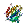
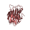
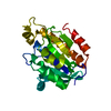
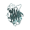
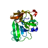
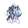
- PDB-5y98: Crystal structure of native unbound peptidyl tRNA hydrolase from ... -
+ Open data

- Basic information
| Entry | Database: PDB / ID: 5y98 | |||||||||
|---|---|---|---|---|---|---|---|---|---|---|
| Title | Crystal structure of native unbound peptidyl tRNA hydrolase from Acinetobacter baumannii at 1.36 A resolution | |||||||||
 Components Components | Peptidyl-tRNA hydrolase Alternative ribosome-rescue factor B Alternative ribosome-rescue factor B | |||||||||
 Keywords Keywords | HYDROLASE | |||||||||
| Function / homology |  Function and homology informationpeptidyl-tRNA hydrolase / aminoacyl-tRNA hydrolase activity / translation / cytoplasm Function and homology informationpeptidyl-tRNA hydrolase / aminoacyl-tRNA hydrolase activity / translation / cytoplasmSimilarity search - Function | |||||||||
| Biological species |  Acinetobacter baumannii (bacteria) Acinetobacter baumannii (bacteria) | |||||||||
| Method | X-RAY DIFFRACTION / SYNCHROTRON / MOLECULAR REPLACEMENT / Resolution: 1.36 Å | |||||||||
 Authors Authors | Iqbal, N. / Singh, N. / Kaushik, S. / Singh, P.K. / Sharma, S. / Singh, T.P. | |||||||||
 Citation Citation | Journal: Biochem. J. / Year: 2018 Title: Search of multiple hot spots on the surface of peptidyl-tRNA hydrolase: structural, binding and antibacterial studies. Authors: Kaushik, S. / Iqbal, N. / Singh, N. / Sikarwar, J.S. / Singh, P.K. / Sharma, P. / Kaur, P. / Sharma, S. / Owais, M. / Singh, T.P. | |||||||||
| History |
|
- Structure visualization
| Structure viewer | Molecule: MolmilJmol/JSmol |
|---|
- Downloads & links
-Download
| PDBx/mmCIF format | 5y98.cif.gz | 99.8 KB | Display | PDBx/mmCIF format |
|---|---|---|---|---|
| PDB format | pdb5y98.ent.gz | 74.1 KB | Display | PDB format |
| PDBx/mmJSON format | 5y98.json.gz | Tree view | PDBx/mmJSON format | |
| Others |  Other downloads Other downloads |
-Validation report
| Arichive directory | https://data.pdbj.org/pub/pdb/validation_reports/y9/5y98ftp://data.pdbj.org/pub/pdb/validation_reports/y9/5y98 | HTTPS FTP |
|---|
-Related structure data
| Related structure data | 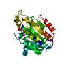 5y9aC  3wh4 S: Starting model for refinement C: citing same article ( |
|---|---|
| Similar structure data |


-Links
 PDBj
PDBj- Assembly
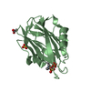
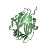
| Deposited unit | 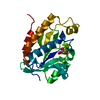
| ||||||||
|---|---|---|---|---|---|---|---|---|---|
| 1 |
| ||||||||
| Unit cell |
|
-Components
| #1: Protein | Alternative ribosome-rescue factor B / PTH Mass: 21250.232 Da / Num. of mol.: 1 Source method: isolated from a genetically manipulated source Source: (gene. exp.) Acinetobacter baumannii (strain ATCC 19606 / DSM 30007 / CIP 70.34 / JCM 6841 / NBRC 109757 / NCIMB 12457 / NCTC 12156 / 81) (bacteria)Strain: ATCC 19606 / DSM 30007 / CIP 70.34 / JCM 6841 / NBRC 109757 / NCIMB 12457 / NCTC 12156 / 81 Gene: pth, F911_03144, HMPREF0010_01329 / Production host: Escherichia coli (E. coli) / References: UniProt: D0C9L6, peptidyl-tRNA hydrolase |
|---|---|
| #2: Chemical | ChemComp-GOL / Glycerol  Mass: 92.094 Da / Num. of mol.: 1 / Source method: obtained synthetically / Formula: C3H8O3 Mass: 92.094 Da / Num. of mol.: 1 / Source method: obtained synthetically / Formula: C3H8O3 |
| #3: Water | ChemComp-HOH / Water Mass: 18.015 Da / Num. of mol.: 216 / Source method: isolated from a natural source / Formula: H2O Mass: 18.015 Da / Num. of mol.: 216 / Source method: isolated from a natural source / Formula: H2O |
-Experimental details
-Experiment
| Experiment | Method: X-RAY DIFFRACTION / Number of used crystals: 1 |
|---|
- Sample preparation
| Crystal | Density Matthews: 2 Å3/Da / Density % sol: 38.53 % |
|---|---|
| Crystal grow | Temperature: 298 K / Method: vapor diffusion, hanging drop / pH: 7.5 Details: 50mM HEPES, PEG 400, PEG 1500, pH 7.5, VAPOR DIFFUSION, HANGING DROP, temperature 298K |
-Data collection
| Diffraction | Mean temperature: 100 K |
|---|---|
| Diffraction source | Source: SYNCHROTRON / Site: ESRF  / Beamline: BM14 / Wavelength: 0.97 Å / Beamline: BM14 / Wavelength: 0.97 Å |
| Detector | Type: MARRESEARCH / Detector: CCD / Date: Jun 23, 2013 / Details: Mirror |
| Radiation | Protocol: SINGLE WAVELENGTH / Monochromatic (M) / Laue (L): M / Scattering type: x-ray |
| Radiation wavelength | Wavelength: 0.97 Å / Relative weight: 1 |
| Reflection | Resolution: 1.36→25.32 Å / Num. obs: 37477 / % possible obs: 99.9 % / Redundancy: 4.2 % / Net I/σ(I): 20.2 |
| Reflection shell | Resolution: 1.36→1.395 Å |
- Processing
| Software |
| ||||||||||||||||||||||||||||||||||||||||||||||||||||||||||||||||||||||||||||||||||||||||||||||||||||||||||||||||||||||||||||||||||||||||||||||||||||||||||||||||||||||||||||||||||||||
|---|---|---|---|---|---|---|---|---|---|---|---|---|---|---|---|---|---|---|---|---|---|---|---|---|---|---|---|---|---|---|---|---|---|---|---|---|---|---|---|---|---|---|---|---|---|---|---|---|---|---|---|---|---|---|---|---|---|---|---|---|---|---|---|---|---|---|---|---|---|---|---|---|---|---|---|---|---|---|---|---|---|---|---|---|---|---|---|---|---|---|---|---|---|---|---|---|---|---|---|---|---|---|---|---|---|---|---|---|---|---|---|---|---|---|---|---|---|---|---|---|---|---|---|---|---|---|---|---|---|---|---|---|---|---|---|---|---|---|---|---|---|---|---|---|---|---|---|---|---|---|---|---|---|---|---|---|---|---|---|---|---|---|---|---|---|---|---|---|---|---|---|---|---|---|---|---|---|---|---|---|---|---|---|
| Refinement | Method to determine structure: MOLECULAR REPLACEMENT Starting model: 3WH4 3wh4 Resolution: 1.36→25.32 Å / Cor.coef. Fo:Fc: 0.983 / Cor.coef. Fo:Fc free: 0.975 / SU B: 1.354 / SU ML: 0.025 / Cross valid method: THROUGHOUT / ESU R: 0.043 / ESU R Free: 0.044 / Details: HYDROGENS HAVE BEEN ADDED IN THE RIDING POSITIONS
| ||||||||||||||||||||||||||||||||||||||||||||||||||||||||||||||||||||||||||||||||||||||||||||||||||||||||||||||||||||||||||||||||||||||||||||||||||||||||||||||||||||||||||||||||||||||
| Solvent computation | Ion probe radii: 0.8 Å / Shrinkage radii: 0.8 Å / VDW probe radii: 1.2 Å | ||||||||||||||||||||||||||||||||||||||||||||||||||||||||||||||||||||||||||||||||||||||||||||||||||||||||||||||||||||||||||||||||||||||||||||||||||||||||||||||||||||||||||||||||||||||
| Displacement parameters | Biso mean: 14.678 Å2
| ||||||||||||||||||||||||||||||||||||||||||||||||||||||||||||||||||||||||||||||||||||||||||||||||||||||||||||||||||||||||||||||||||||||||||||||||||||||||||||||||||||||||||||||||||||||
| Refinement step | Cycle: 1 / Resolution: 1.36→25.32 Å
| ||||||||||||||||||||||||||||||||||||||||||||||||||||||||||||||||||||||||||||||||||||||||||||||||||||||||||||||||||||||||||||||||||||||||||||||||||||||||||||||||||||||||||||||||||||||
| Refine LS restraints |
|