 Movie
Movie Controller


+ Open data

- Basic information
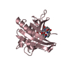
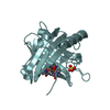
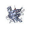
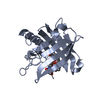
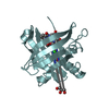
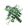
| Entry | Database: PDB / ID: 3dsz | ||||||
|---|---|---|---|---|---|---|---|
| Title | Engineered human lipocalin 2 in complex with Y-DTPA | ||||||
 Components Components | engineered human lipocalin 2 | ||||||
 Keywords Keywords |  TRANSPORT PROTEIN / protein design / ligand binding protein / beta barrel / engineered lipocalin / de novo protein / protein binding TRANSPORT PROTEIN / protein design / ligand binding protein / beta barrel / engineered lipocalin / de novo protein / protein binding | ||||||
| Function / homology |  Function and homology information Function and homology informationsiderophore transport / Metal sequestration by antimicrobial proteins / iron ion sequestering activity / enterobactin binding / Iron uptake and transport / specific granule lumen / positive regulation of cold-induced thermogenesis / Interleukin-4 and Interleukin-13 signaling / defense response to bacterium / iron ion binding ...siderophore transport / Metal sequestration by antimicrobial proteins / iron ion sequestering activity / enterobactin binding / Iron uptake and transport / specific granule lumen / positive regulation of cold-induced thermogenesis / Interleukin-4 and Interleukin-13 signaling / defense response to bacterium / iron ion binding / innate immune response / apoptotic process / Neutrophil degranulation / extracellular space / extracellular exosome / extracellular region / identical protein bindingSimilarity search - Function | ||||||
| Biological species |  Homo sapiens (human) Homo sapiens (human) | ||||||
| Method | X-RAY DIFFRACTION / SYNCHROTRON / MOLECULAR REPLACEMENT / molecular replacement / Resolution: 2 Å | ||||||
 Authors Authors | Eichinger, A. / Skerra, A. | ||||||
 Citation Citation | Journal: J.Am.Chem.Soc. / Year: 2009 Title: High-affinity recognition of lanthanide(III) chelate complexes by a reprogrammed human lipocalin 2 Authors: Kim, H.J. / Eichinger, A. / Skerra, A. | ||||||
| History |
|
- Structure visualization
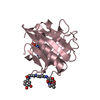
| Structure viewer | Molecule: MolmilJmol/JSmol |
|---|
- Downloads & links
-Download
| PDBx/mmCIF format | 3dsz.cif.gz | 87.7 KB | Display | PDBx/mmCIF format |
|---|---|---|---|---|
| PDB format | pdb3dsz.ent.gz | 72 KB | Display | PDB format |
| PDBx/mmJSON format | 3dsz.json.gz | Tree view | PDBx/mmJSON format | |
| Others |  Other downloads Other downloads |
-Validation report
| Arichive directory | https://data.pdbj.org/pub/pdb/validation_reports/ds/3dszftp://data.pdbj.org/pub/pdb/validation_reports/ds/3dsz | HTTPS FTP |
|---|
-Related structure data


-Links
 PDBj
PDBj


- Assembly
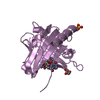
| Deposited unit | 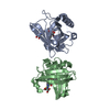
| ||||||||
|---|---|---|---|---|---|---|---|---|---|
| 1 | 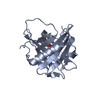
| ||||||||
| 2 | 
| ||||||||
| Unit cell |
|
-Components
| #1: Protein | Mass: 21344.191 Da / Num. of mol.: 2 Source method: isolated from a genetically manipulated source Source: (gene. exp.) Homo sapiens (human) / Gene: LCN2, variant Tb7.N9 / Plasmid: pNGAL15 / Production host:  Escherichia coli (E. coli) / Strain (production host): BL21 / References: UniProt: P80188*PLUS Escherichia coli (E. coli) / Strain (production host): BL21 / References: UniProt: P80188*PLUS#2: Chemical | 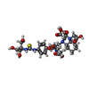  Mass: 715.769 Da / Num. of mol.: 2 / Source method: obtained synthetically / Formula: C30H45N5O13S Mass: 715.769 Da / Num. of mol.: 2 / Source method: obtained synthetically / Formula: C30H45N5O13S#3: Chemical | Yttrium  Mass: 88.906 Da / Num. of mol.: 2 / Source method: obtained synthetically / Formula: Y Mass: 88.906 Da / Num. of mol.: 2 / Source method: obtained synthetically / Formula: Y#4: Water | ChemComp-HOH / | Water Mass: 18.015 Da / Num. of mol.: 328 / Source method: isolated from a natural source / Formula: H2O Mass: 18.015 Da / Num. of mol.: 328 / Source method: isolated from a natural source / Formula: H2OSequence details | A SEQUENCE DATABASE REFERENCE FOR THIS PROTEIN DOES NOT CURRENTLY EXIST. THIS SEQUENCE WILL BE ...A SEQUENCE DATABASE REFERENCE FOR THIS PROTEIN DOES NOT CURRENTLY EXIST. THIS SEQUENCE WILL BE DEPOSITED IN THE SEQUENCE DATABASE. | |
|---|
-Experimental details
-Experiment
| Experiment | Method: X-RAY DIFFRACTION / Number of used crystals: 1 |
|---|
- Sample preparation
| Crystal | Density Matthews: 2.29 Å3/Da / Density % sol: 46.2 % |
|---|---|
| Crystal grow | Temperature: 293 K / Method: vapor diffusion, hanging drop / pH: 5.5 Details: 22% polyethylene glycol 3350, 0.1M Bistris/HCl, pH5.5, VAPOR DIFFUSION, HANGING DROP, temperature 293K |
-Data collection
| Diffraction | Mean temperature: 100 K |
|---|---|
| Diffraction source | Source: SYNCHROTRON / Site: BESSY  / Beamline: 14.1 / Wavelength: 0.95373 Å / Beamline: 14.1 / Wavelength: 0.95373 Å |
| Detector | Type: MARMOSAIC 225 mm CCD / Detector: CCD / Date: Mar 10, 2007 / Details: mirrors |
| Radiation | Monochromator: Si 111 CRYSTAL / Protocol: SINGLE WAVELENGTH / Monochromatic (M) / Laue (L): M / Scattering type: x-ray |
| Radiation wavelength | Wavelength: 0.95373 Å / Relative weight: 1 |
| Reflection | Resolution: 2→66.979 Å / Num. obs: 26827 / % possible obs: 98.2 % / Redundancy: 9.7 % / Rmerge(I) obs: 0.109 / Net I/σ(I): 4.1 |
| Reflection shell | Resolution: 2→2.11 Å / Redundancy: 9.8 % / Rmerge(I) obs: 0.372 / Mean I/σ(I) obs: 6.1 / Num. unique all: 3813 / Rsym value: 0.372 / % possible all: 97.4 |
-Phasing
| Phasing | Method: molecular replacement | |||||||||
|---|---|---|---|---|---|---|---|---|---|---|
| Phasing MR | Rfactor: 0.423 / Cor.coef. Fo:Fc: 0.579
|
- Processing
| Software |
| |||||||||||||||||||||||||
|---|---|---|---|---|---|---|---|---|---|---|---|---|---|---|---|---|---|---|---|---|---|---|---|---|---|---|
| Refinement | Method to determine structure: MOLECULAR REPLACEMENT / Resolution: 2→40 Å / Occupancy max: 1 / Occupancy min: 0.01 / FOM work R set: 0.864 / σ(F): 0 / Stereochemistry target values: Engh & Huber
| |||||||||||||||||||||||||
| Solvent computation | Bsol: 56.905 Å2 | |||||||||||||||||||||||||
| Displacement parameters | Biso max: 81.5 Å2 / Biso mean: 27.759 Å2 / Biso min: 11.34 Å2
| |||||||||||||||||||||||||
| Refinement step | Cycle: LAST / Resolution: 2→40 Å
| |||||||||||||||||||||||||
| Refine LS restraints |
| |||||||||||||||||||||||||
| Xplor file |
|
