 Movie
Movie Controller

[English] 日本語
 Yorodumi
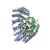
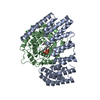
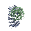
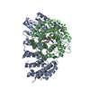
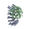
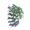
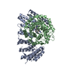
- PDB-3dsx: Crystal structure of RabGGTase(DELTA LRR; DELTA IG)in complex wit... -
+ Open data

- Basic information
| Entry | Database: PDB / ID: 3dsx | ||||||
|---|---|---|---|---|---|---|---|
| Title | Crystal structure of RabGGTase(DELTA LRR; DELTA IG)in complex with di-prenylated peptide Ser-Cys(GG)-Ser-Cys(GG) derivated from Rab7 | ||||||
 Components Components | (Geranylgeranyl transferase type-2 subunit ...) x 2 | ||||||
 Keywords Keywords |  TRANSFERASE / protein prenylation / Metal-binding / Prenyltransferase / Zinc / Phosphoprotein TRANSFERASE / protein prenylation / Metal-binding / Prenyltransferase / Zinc / Phosphoprotein | ||||||
| Function / homology |  Function and homology informationisoprenoid binding / TP53 regulates transcription of several additional cell death genes whose specific roles in p53-dependent apoptosis remain uncertain / protein geranylgeranyltransferase type II / RAB geranylgeranylation / Rab-protein geranylgeranyltransferase complex / Rab geranylgeranyltransferase activity / protein geranylgeranylation / small GTPase binding / zinc ion binding / cytoplasm Function and homology informationisoprenoid binding / TP53 regulates transcription of several additional cell death genes whose specific roles in p53-dependent apoptosis remain uncertain / protein geranylgeranyltransferase type II / RAB geranylgeranylation / Rab-protein geranylgeranyltransferase complex / Rab geranylgeranyltransferase activity / protein geranylgeranylation / small GTPase binding / zinc ion binding / cytoplasmSimilarity search - Function | ||||||
| Biological species |  Rattus norvegicus (Norway rat) Rattus norvegicus (Norway rat) | ||||||
| Method | X-RAY DIFFRACTION / SYNCHROTRON / MOLECULAR REPLACEMENT / Resolution: 2.1 Å | ||||||
 Authors Authors | Guo, Z. / Yu, S. / Goody, R.S. / Alexandrov, K. / Blankenfeldt, W. | ||||||
 Citation Citation | Journal: Embo J. / Year: 2008 Title: Structures of RabGGTase-substrate/product complexes provide insights into the evolution of protein prenylation Authors: Guo, Z. / Wu, Y.-W. / Das, D. / Delon, C. / Cramer, J. / Yu, S. / Thuns, S. / Lupilova, N. / Waldmann, H. / Brunsveld, L. / Goody, R.S. / Alexandrov, K. / Blankenfeldt, W. | ||||||
| History |
|
- Structure visualization
| Structure viewer | Molecule: MolmilJmol/JSmol |
|---|
- Downloads & links
-Download
| PDBx/mmCIF format | 3dsx.cif.gz | 272.3 KB | Display | PDBx/mmCIF format |
|---|---|---|---|---|
| PDB format | pdb3dsx.ent.gz | 218.4 KB | Display | PDB format |
| PDBx/mmJSON format | 3dsx.json.gz | Tree view | PDBx/mmJSON format | |
| Others |  Other downloads Other downloads |
-Validation report
| Arichive directory | https://data.pdbj.org/pub/pdb/validation_reports/ds/3dsxftp://data.pdbj.org/pub/pdb/validation_reports/ds/3dsx | HTTPS FTP |
|---|
-Related structure data
| Related structure data |  3dssSC  3dstC 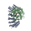 3dsuC 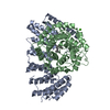 3dsvC 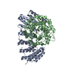 3dswC S: Starting model for refinement C: citing same article ( |
|---|---|
| Similar structure data |


-Links
 PDBj
PDBj

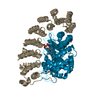
- Assembly
| Deposited unit | 
| ||||||||
|---|---|---|---|---|---|---|---|---|---|
| 1 |
| ||||||||
| Unit cell |
|
-Components
-Geranylgeranyl transferase type-2 subunit ... , 2 types, 2 molecules AB
| #1: Protein | Mass: 38441.598 Da / Num. of mol.: 1 / Fragment: PFTA domains, UNP residues 1-237 and 353-441 Source method: isolated from a genetically manipulated source Source: (gene. exp.) Rattus norvegicus (Norway rat)Description: coexpression of engineered alpha-subunit from pGATEV and beta-subunit from pET3 0a Gene: Rabggta, Ggta / Plasmid: pGATEV and pET30a / Production host:  Escherichia coli (E. coli) / Strain (production host): BL21 CODON-PLUS RIL (DE3) Escherichia coli (E. coli) / Strain (production host): BL21 CODON-PLUS RIL (DE3)References: UniProt: Q08602, protein geranylgeranyltransferase type II |
|---|---|
| #2: Protein | Mass: 36892.160 Da / Num. of mol.: 1 Source method: isolated from a genetically manipulated source Source: (gene. exp.) Rattus norvegicus (Norway rat)Description: coexpression of engineered alpha-subunit from pGATEV and beta-subunit from pET3 0a Gene: Rabggtb, Ggtb / Plasmid: pGATEV and pET30a / Production host: Escherichia coli (E. coli) / Strain (production host): BL21 CODON-PLUS RIL (DE3)References: UniProt: Q08603, protein geranylgeranyltransferase type II |
-Non-polymers , 4 types, 300 molecules 


| #3: Chemical | ChemComp-ZN /  Mass: 65.409 Da / Num. of mol.: 1 / Source method: obtained synthetically / Formula: Zn Mass: 65.409 Da / Num. of mol.: 1 / Source method: obtained synthetically / Formula: Zn |
|---|---|
| #4: Chemical | ChemComp-CA /  Mass: 40.078 Da / Num. of mol.: 1 / Source method: obtained synthetically / Formula: Ca Mass: 40.078 Da / Num. of mol.: 1 / Source method: obtained synthetically / Formula: Ca |
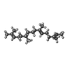
| #5: Chemical | ChemComp-GER /  Mass: 274.484 Da / Num. of mol.: 1 / Source method: obtained synthetically / Formula: C20H34 Mass: 274.484 Da / Num. of mol.: 1 / Source method: obtained synthetically / Formula: C20H34 |
| #6: Water | ChemComp-HOH / WaterMass: 18.015 Da / Num. of mol.: 297 / Source method: isolated from a natural source / Formula: H2O |
-Details
| Nonpolymer details | THE DEPOSITORS HAVE SOAKED THE CRYSTALS WITH THE DIPRENYLATED PEPTIDE, SER-CYS(GG)-SER-CYS(GG), BUT ...THE DEPOSITORS |
|---|
-Experimental details
-Experiment
| Experiment | Method: X-RAY DIFFRACTION / Number of used crystals: 1 |
|---|
- Sample preparation
| Crystal | Density Matthews: 2.33 Å3/Da / Density % sol: 47.32 % |
|---|---|
| Crystal grow | Temperature: 284 K / Method: vapor diffusion, hanging drop / pH: 7.2 Details: 14% (w/v) PEG 3350, 0.2M Ca(OAc)2, 2.5% (v/v)DMSO, 0.1M HEPES, pH 7.2, VAPOR DIFFUSION, HANGING DROP, temperature 284K |
-Data collection
| Diffraction | Mean temperature: 100 K |
|---|---|
| Diffraction source | Source: SYNCHROTRON / Site: SLS  / Beamline: X10SA / Wavelength: 0.9841 Å / Beamline: X10SA / Wavelength: 0.9841 Å |
| Detector | Type: MARMOSAIC 225 mm CCD / Detector: CCD / Date: Oct 27, 2007 |
| Radiation | Monochromator: SI(111) / Protocol: SINGLE WAVELENGTH / Monochromatic (M) / Laue (L): M / Scattering type: x-ray |
| Radiation wavelength | Wavelength: 0.9841 Å / Relative weight: 1 |
| Reflection | Resolution: 2.1→30 Å / Num. all: 41909 / Num. obs: 41826 / % possible obs: 99.8 % / Observed criterion σ(I): 5 / Redundancy: 7.2 % / Biso Wilson estimate: 39 Å2 / Rsym value: 0.057 / Net I/σ(I): 23.4 |
| Reflection shell | Resolution: 2.1→2.2 Å / Redundancy: 7.4 % / Mean I/σ(I) obs: 6.9 / Num. unique all: 5348 / Rsym value: 0.391 / % possible all: 100 |
- Processing
| Software |
| ||||||||||||||||||||||||||||||||||||||||||||||||||||||||||||||||||||||||||||||||||||||||||||||||||||||||||||||||||||||||||||||||||||||||||||||||||||||||||||||||||||||||||||||||||||||||||||||||||||||||
|---|---|---|---|---|---|---|---|---|---|---|---|---|---|---|---|---|---|---|---|---|---|---|---|---|---|---|---|---|---|---|---|---|---|---|---|---|---|---|---|---|---|---|---|---|---|---|---|---|---|---|---|---|---|---|---|---|---|---|---|---|---|---|---|---|---|---|---|---|---|---|---|---|---|---|---|---|---|---|---|---|---|---|---|---|---|---|---|---|---|---|---|---|---|---|---|---|---|---|---|---|---|---|---|---|---|---|---|---|---|---|---|---|---|---|---|---|---|---|---|---|---|---|---|---|---|---|---|---|---|---|---|---|---|---|---|---|---|---|---|---|---|---|---|---|---|---|---|---|---|---|---|---|---|---|---|---|---|---|---|---|---|---|---|---|---|---|---|---|---|---|---|---|---|---|---|---|---|---|---|---|---|---|---|---|---|---|---|---|---|---|---|---|---|---|---|---|---|---|---|---|---|
| Refinement | Method to determine structure: MOLECULAR REPLACEMENT Starting model: DB ENTRY 3DSS Resolution: 2.1→29.37 Å / Cor.coef. Fo:Fc: 0.961 / Cor.coef. Fo:Fc free: 0.923 / SU B: 11.084 / SU ML: 0.135 / Cross valid method: THROUGHOUT / ESU R Free: 0.181 / Stereochemistry target values: MAXIMUM LIKELIHOOD / Details: HYDROGENS HAVE BEEN ADDED IN THE RIDING POSITIONS
| ||||||||||||||||||||||||||||||||||||||||||||||||||||||||||||||||||||||||||||||||||||||||||||||||||||||||||||||||||||||||||||||||||||||||||||||||||||||||||||||||||||||||||||||||||||||||||||||||||||||||
| Solvent computation | Ion probe radii: 0.8 Å / Shrinkage radii: 0.8 Å / VDW probe radii: 1.2 Å / Solvent model: BABINET MODEL WITH MASK | ||||||||||||||||||||||||||||||||||||||||||||||||||||||||||||||||||||||||||||||||||||||||||||||||||||||||||||||||||||||||||||||||||||||||||||||||||||||||||||||||||||||||||||||||||||||||||||||||||||||||
| Displacement parameters | Biso mean: 30.065 Å2 | ||||||||||||||||||||||||||||||||||||||||||||||||||||||||||||||||||||||||||||||||||||||||||||||||||||||||||||||||||||||||||||||||||||||||||||||||||||||||||||||||||||||||||||||||||||||||||||||||||||||||
| Refinement step | Cycle: LAST / Resolution: 2.1→29.37 Å
| ||||||||||||||||||||||||||||||||||||||||||||||||||||||||||||||||||||||||||||||||||||||||||||||||||||||||||||||||||||||||||||||||||||||||||||||||||||||||||||||||||||||||||||||||||||||||||||||||||||||||
| Refine LS restraints |
| ||||||||||||||||||||||||||||||||||||||||||||||||||||||||||||||||||||||||||||||||||||||||||||||||||||||||||||||||||||||||||||||||||||||||||||||||||||||||||||||||||||||||||||||||||||||||||||||||||||||||
| LS refinement shell | Resolution: 2.1→2.154 Å / Total num. of bins used: 20
| ||||||||||||||||||||||||||||||||||||||||||||||||||||||||||||||||||||||||||||||||||||||||||||||||||||||||||||||||||||||||||||||||||||||||||||||||||||||||||||||||||||||||||||||||||||||||||||||||||||||||
| Refinement TLS params. | Method: refined / Refine-ID: X-RAY DIFFRACTION
| ||||||||||||||||||||||||||||||||||||||||||||||||||||||||||||||||||||||||||||||||||||||||||||||||||||||||||||||||||||||||||||||||||||||||||||||||||||||||||||||||||||||||||||||||||||||||||||||||||||||||
| Refinement TLS group |
|
