 Movie
Movie Controller


+ Open data

- Basic information
| Entry | Database: PDB / ID: 3ds7 | ||||||
|---|---|---|---|---|---|---|---|
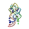
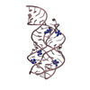
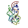
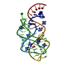
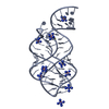
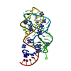
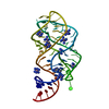
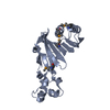
| Title | Structure of an RNA-2'-deoxyguanosine complex | ||||||
 Components Components | 67-MER | ||||||
 Keywords Keywords |  RNA / RNA-ligand complex / riboswitch RNA / RNA-ligand complex / riboswitch | ||||||
| Function / homology | ACETATE ION / 2'-DEOXY-GUANOSINE / COBALT HEXAMMINE(III) / RNA / RNA (> 10) Function and homology information Function and homology information | ||||||
| Method | X-RAY DIFFRACTION / MOLECULAR REPLACEMENT / Resolution: 1.85 Å | ||||||
 Authors Authors | Edwards, A.L. / Batey, R.T. | ||||||
 Citation Citation | Journal: J.Mol.Biol. / Year: 2009 Title: A structural basis for the recognition of 2'-deoxyguanosine by the purine riboswitch. Authors: Edwards, A.L. / Batey, R.T. | ||||||
| History |
|
- Structure visualization
| Structure viewer | Molecule: MolmilJmol/JSmol |
|---|
- Downloads & links
-Download
| PDBx/mmCIF format | 3ds7.cif.gz | 104.1 KB | Display | PDBx/mmCIF format |
|---|---|---|---|---|
| PDB format | pdb3ds7.ent.gz | 76.7 KB | Display | PDB format |
| PDBx/mmJSON format | 3ds7.json.gz | Tree view | PDBx/mmJSON format | |
| Others |  Other downloads Other downloads |
-Validation report
| Arichive directory | https://data.pdbj.org/pub/pdb/validation_reports/ds/3ds7ftp://data.pdbj.org/pub/pdb/validation_reports/ds/3ds7 | HTTPS FTP |
|---|
-Related structure data
| Related structure data |  1u8d S: Starting model for refinement |
|---|---|
| Similar structure data |


-Links
 PDBj
PDBj


- Assembly
| Deposited unit | 
| ||||||||
|---|---|---|---|---|---|---|---|---|---|
| 1 | 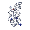
| ||||||||
| 2 | 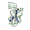
| ||||||||
| Unit cell |
|
-Components
| #1: RNA chain | Mass: 21542.873 Da / Num. of mol.: 2 / Source method: obtained synthetically Details: This sequence is synthetic derived from a mutagenesis study #2: Chemical | Acetate  Mass: 59.044 Da / Num. of mol.: 2 / Source method: obtained synthetically / Formula: C2H3O2 Mass: 59.044 Da / Num. of mol.: 2 / Source method: obtained synthetically / Formula: C2H3O2#3: Chemical | ChemComp-NCO /   Mass: 161.116 Da / Num. of mol.: 11 / Source method: obtained synthetically / Formula: CoH18N6 Mass: 161.116 Da / Num. of mol.: 11 / Source method: obtained synthetically / Formula: CoH18N6#4: Chemical | Deoxyguanosine  Mass: 267.241 Da / Num. of mol.: 2 / Source method: obtained synthetically / Formula: C10H13N5O4 Mass: 267.241 Da / Num. of mol.: 2 / Source method: obtained synthetically / Formula: C10H13N5O4#5: Water | ChemComp-HOH / | Water Mass: 18.015 Da / Num. of mol.: 622 / Source method: isolated from a natural source / Formula: H2O Mass: 18.015 Da / Num. of mol.: 622 / Source method: isolated from a natural source / Formula: H2O |
|---|
-Experimental details
-Experiment
| Experiment | Method: X-RAY DIFFRACTION / Number of used crystals: 1 |
|---|
- Sample preparation
| Crystal | Density Matthews: 2.18 Å3/Da / Density % sol: 43.6 % | ||||||||||||||||||||||||||||||||||||||||||||
|---|---|---|---|---|---|---|---|---|---|---|---|---|---|---|---|---|---|---|---|---|---|---|---|---|---|---|---|---|---|---|---|---|---|---|---|---|---|---|---|---|---|---|---|---|---|
| Crystal grow | Temperature: 298 K / Method: vapor diffusion, hanging drop / pH: 7.5 Details: 0.01M K-HEPES, 0.0119 M cobalt(III) hexammine, 22% PEG 2000, 0.66M ammonium acetate, pH 7.5, VAPOR DIFFUSION, HANGING DROP, temperature 298K | ||||||||||||||||||||||||||||||||||||||||||||
| Components of the solutions |
|
-Data collection
| Diffraction | Mean temperature: 100 K |
|---|---|
| Diffraction source | Source: ROTATING ANODE / Type: RIGAKU RU200 / Wavelength: 1.54 Å |
| Detector | Type: RIGAKU RAXIS IV++ / Detector: IMAGE PLATE / Date: Nov 12, 2007 |
| Radiation | Monochromator: Ni Filter / Protocol: SINGLE WAVELENGTH / Monochromatic (M) / Laue (L): M / Scattering type: x-ray |
| Radiation wavelength | Wavelength: 1.54 Å / Relative weight: 1 |
| Reflection | Resolution: 1.85→19.5 Å / Num. all: 31039 / Num. obs: 29177 / % possible obs: 94 % / Observed criterion σ(I): 2 / Redundancy: 3.56 % / Biso Wilson estimate: 20 Å2 / Rmerge(I) obs: 0.075 / Net I/σ(I): 8.4 |
| Reflection shell | Resolution: 1.85→1.92 Å / Redundancy: 3.57 % / Rmerge(I) obs: 0.297 / Mean I/σ(I) obs: 3.4 / Num. unique all: 2822 / % possible all: 91.4 |
- Processing
| Software |
| |||||||||||||||||||||||||
|---|---|---|---|---|---|---|---|---|---|---|---|---|---|---|---|---|---|---|---|---|---|---|---|---|---|---|
| Refinement | Method to determine structure: MOLECULAR REPLACEMENT Starting model: PDB 1U8D 1u8d Resolution: 1.85→19.5 Å / Rfactor Rfree error: 0.005 / Data cutoff high absF: 518064.88 / Data cutoff low absF: 0 / Isotropic thermal model: RESTRAINED / Cross valid method: THROUGHOUT / σ(F): 0 / σ(I): 2 / Stereochemistry target values: Engh & Huber / Details: BULK SOLVENT MODEL USED
| |||||||||||||||||||||||||
| Solvent computation | Solvent model: FLAT MODEL / Bsol: 74.7773 Å2 / ksol: 0.45 e/Å3 | |||||||||||||||||||||||||
| Displacement parameters | Biso mean: 34.4 Å2
| |||||||||||||||||||||||||
| Refine analyze |
| |||||||||||||||||||||||||
| Refinement step | Cycle: LAST / Resolution: 1.85→19.5 Å
| |||||||||||||||||||||||||
| Refine LS restraints |
| |||||||||||||||||||||||||
| LS refinement shell | Resolution: 1.85→1.97 Å / Rfactor Rfree error: 0.015 / Total num. of bins used: 6
| |||||||||||||||||||||||||
| Xplor file |
|
