 Movie
Movie Controller

[English] 日本語
 Yorodumi
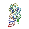
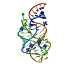
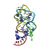
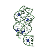
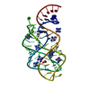
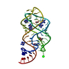
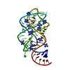
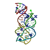
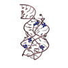
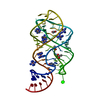
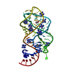
- PDB-4feo: Crystal structure of the AU25A/A46G/C74U mutant xpt-pbuX guanine ... -
+ Open data

- Basic information
| Entry | Database: PDB / ID: 4feo | ||||||
|---|---|---|---|---|---|---|---|
| Title | Crystal structure of the AU25A/A46G/C74U mutant xpt-pbuX guanine riboswitch aptamer domain in complex with 2,6-diaminopurine | ||||||
 Components Components | U25A/A46G/C74U mutant of the B. subtilis xpt-pbuX guanine riboswitch aptamer domain | ||||||
 Keywords Keywords |  RNA / three-way junction with distal tertiary interaction / genetic regulatory element / 2 / 6-diaminopurine RNA / three-way junction with distal tertiary interaction / genetic regulatory element / 2 / 6-diaminopurine | ||||||
| Function / homology | 9H-PURINE-2,6-DIAMINE / COBALT HEXAMMINE(III) / RNA / RNA (> 10) Function and homology information Function and homology information | ||||||
| Method | X-RAY DIFFRACTION / MOLECULAR REPLACEMENT / Resolution: 1.6 Å | ||||||
 Authors Authors | Stoddard, C.D. / Trausch, J.J. / Widmann, J. / Marcano, J. / Knight, R. / Batey, R.T. | ||||||
 Citation Citation | Journal: J.Mol.Biol. / Year: 2013 Title: Nucleotides Adjacent to the Ligand-Binding Pocket are Linked to Activity Tuning in the Purine Riboswitch. Authors: Stoddard, C.D. / Widmann, J. / Trausch, J.J. / Marcano-Velazquez, J.G. / Knight, R. / Batey, R.T. | ||||||
| History |
|
- Structure visualization
| Structure viewer | Molecule: MolmilJmol/JSmol |
|---|
- Downloads & links
-Download
| PDBx/mmCIF format | 4feo.cif.gz | 66 KB | Display | PDBx/mmCIF format |
|---|---|---|---|---|
| PDB format | pdb4feo.ent.gz | 47.3 KB | Display | PDB format |
| PDBx/mmJSON format | 4feo.json.gz | Tree view | PDBx/mmJSON format | |
| Others |  Other downloads Other downloads |
-Validation report
| Arichive directory | https://data.pdbj.org/pub/pdb/validation_reports/fe/4feoftp://data.pdbj.org/pub/pdb/validation_reports/fe/4feo | HTTPS FTP |
|---|
-Related structure data


-Links
 PDBj
PDBj


- Assembly
| Deposited unit | 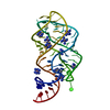
| ||||||||
|---|---|---|---|---|---|---|---|---|---|
| 1 |
| ||||||||
| Unit cell |
|
-Components
| #1: RNA chain | Mass: 21546.809 Da / Num. of mol.: 1 / Source method: obtained synthetically / Details: T7 RNA transcript | ||
|---|---|---|---|
| #2: Chemical | ChemComp-6AP / 2,6-Diaminopurine  Mass: 150.141 Da / Num. of mol.: 1 / Source method: obtained synthetically / Formula: C5H6N6 Mass: 150.141 Da / Num. of mol.: 1 / Source method: obtained synthetically / Formula: C5H6N6 | ||
| #3: Chemical | ChemComp-NCO /   Mass: 161.116 Da / Num. of mol.: 9 / Source method: obtained synthetically / Formula: CoH18N6 Mass: 161.116 Da / Num. of mol.: 9 / Source method: obtained synthetically / Formula: CoH18N6#4: Water | ChemComp-HOH / | Water Mass: 18.015 Da / Num. of mol.: 689 / Source method: isolated from a natural source / Formula: H2O Mass: 18.015 Da / Num. of mol.: 689 / Source method: isolated from a natural source / Formula: H2O |
-Experimental details
-Experiment
| Experiment | Method: X-RAY DIFFRACTION / Number of used crystals: 1 |
|---|
- Sample preparation
| Crystal | Density Matthews: 2.25 Å3/Da / Density % sol: 45.39 % |
|---|---|
| Crystal grow | Temperature: 303 K / Method: vapor diffusion, hanging drop / pH: 7.5 Details: 25% PEG 3K, 200 mM ammonium acetate, 10 mM cobalt hexammine, pH 7.5, VAPOR DIFFUSION, HANGING DROP, temperature 303K |
-Data collection
| Diffraction | Mean temperature: 100 K |
|---|---|
| Diffraction source | Source: ROTATING ANODE / Type: RIGAKU / Wavelength: 1.5418 Å |
| Detector | Type: RIGAKU RAXIS IV / Detector: IMAGE PLATE / Date: May 30, 2009 / Details: OSMIC |
| Radiation | Monochromator: NICKEL FILTER / Protocol: SINGLE WAVELENGTH / Monochromatic (M) / Laue (L): M / Scattering type: x-ray |
| Radiation wavelength | Wavelength: 1.5418 Å / Relative weight: 1 |
| Reflection | Resolution: 1.6→20 Å / Num. all: 25620 / Num. obs: 24953 / % possible obs: 98.6 % / Observed criterion σ(F): 2 / Observed criterion σ(I): 2 |
| Reflection shell | Resolution: 1.6→1.66 Å / % possible all: 98.6 |
- Processing
| Software |
| ||||||||||||||||||||
|---|---|---|---|---|---|---|---|---|---|---|---|---|---|---|---|---|---|---|---|---|---|
| Refinement | Method to determine structure: MOLECULAR REPLACEMENT / Resolution: 1.6→20 Å / σ(F): 2 / Stereochemistry target values: Engh & Huber
| ||||||||||||||||||||
| Refine analyze | Luzzati coordinate error obs: 0.18 Å / Luzzati d res low obs: 5 Å / Luzzati sigma a obs: 0.15 Å | ||||||||||||||||||||
| Refinement step | Cycle: LAST / Resolution: 1.6→20 Å
| ||||||||||||||||||||
| Refine LS restraints |
|
