 Movie
Movie Controller


+ Open data

- Basic information
| Entry | Database: PDB / ID: 3dmm | ||||||
|---|---|---|---|---|---|---|---|
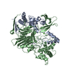
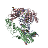
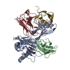
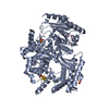
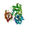
| Title | Crystal structure of the CD8 alpha beta/H-2Dd complex | ||||||
 Components Components |
| ||||||
 Keywords Keywords |  IMMUNE SYSTEM / T cell co-receptor CD8ab MHC complex / Glycoprotein / Immune response / Membrane / MHC I / Phosphoprotein / Transmembrane / Immunoglobulin domain / Secreted / Envelope protein / Alternative splicing / Polymorphism IMMUNE SYSTEM / T cell co-receptor CD8ab MHC complex / Glycoprotein / Immune response / Membrane / MHC I / Phosphoprotein / Transmembrane / Immunoglobulin domain / Secreted / Envelope protein / Alternative splicing / Polymorphism | ||||||
| Function / homology |  Function and homology information Function and homology informationcytotoxic T cell differentiation / MHC class I protein complex binding / T cell mediated immunity / Endosomal/Vacuolar pathway / DAP12 interactions / Antigen Presentation: Folding, assembly and peptide loading of class I MHC / ER-Phagosome pathway / DAP12 signaling / Immunoregulatory interactions between a Lymphoid and a non-Lymphoid cell / regulation of membrane depolarization ...cytotoxic T cell differentiation / MHC class I protein complex binding / T cell mediated immunity / Endosomal/Vacuolar pathway / DAP12 interactions / Antigen Presentation: Folding, assembly and peptide loading of class I MHC / ER-Phagosome pathway / DAP12 signaling / Immunoregulatory interactions between a Lymphoid and a non-Lymphoid cell / regulation of membrane depolarization / MHC class I protein binding / plasma membrane raft / antigen processing and presentation of endogenous peptide antigen via MHC class I via ER pathway, TAP-dependent / cellular defense response / beta-2-microglobulin binding / coreceptor activity / positive regulation of calcium-mediated signaling / T cell activation / Neutrophil degranulation / antigen processing and presentation of endogenous peptide antigen via MHC class I via ER pathway, TAP-independent / antigen processing and presentation of endogenous peptide antigen via MHC class Ib / calcium-mediated signaling / lumenal side of endoplasmic reticulum membrane / peptide binding / cellular response to iron(III) ion / antigen processing and presentation of exogenous protein antigen via MHC class Ib, TAP-dependent / negative regulation of forebrain neuron differentiation / regulation of erythrocyte differentiation / peptide antigen assembly with MHC class I protein complex / response to molecule of bacterial origin / regulation of iron ion transport / MHC class I peptide loading complex / HFE-transferrin receptor complex / T cell mediated cytotoxicity / cellular response to iron ion / antigen processing and presentation of endogenous peptide antigen via MHC class I / positive regulation of T cell cytokine production / MHC class I protein complex / multicellular organismal-level iron ion homeostasis / negative regulation of neurogenesis / peptide antigen assembly with MHC class II protein complex / positive regulation of receptor-mediated endocytosis / MHC class II protein complex / cellular response to nicotine / positive regulation of T cell mediated cytotoxicity / phagocytic vesicle membrane / peptide antigen binding / positive regulation of cellular senescence / antigen processing and presentation of exogenous peptide antigen via MHC class II / negative regulation of epithelial cell proliferation / positive regulation of immune response / antimicrobial humoral immune response mediated by antimicrobial peptide / sensory perception of smell / positive regulation of T cell activation / negative regulation of neuron projection development / MHC class II protein complex binding / late endosome membrane / T cell differentiation in thymus / T cell receptor signaling pathway / iron ion transport / protein refolding / antibacterial humoral response / protein homotetramerization / defense response to virus / intracellular iron ion homeostasis / adaptive immune response / cellular response to lipopolysaccharide / defense response to Gram-negative bacterium / amyloid fibril formation / learning or memory / cell surface receptor signaling pathway / receptor complex / defense response to Gram-positive bacterium / immune response / lysosomal membrane / external side of plasma membrane / signaling receptor binding / innate immune response / protein-containing complex binding / protein kinase binding / structural molecule activity / Golgi apparatus / cell surface / protein homodimerization activity / extracellular space / identical protein binding / plasma membrane / cytosolSimilarity search - Function | ||||||
| Biological species |  Mus musculus (house mouse) Mus musculus (house mouse) | ||||||
| Method | X-RAY DIFFRACTION / SYNCHROTRON / MOLECULAR REPLACEMENT / Resolution: 2.6 Å | ||||||
 Authors Authors | Wang, R. / Natarajan, K. / Margulies, D.H. | ||||||
 Citation Citation | Journal: J.Immunol. / Year: 2009 Title: Structural basis of the CD8alphabeta/MHC class i interaction: focused recognition orients CD8beta to a T cell proximal position. Authors: Wang, R. / Natarajan, K. / Margulies, D.H. | ||||||
| History |
|
- Structure visualization
| Structure viewer | Molecule: MolmilJmol/JSmol |
|---|
- Downloads & links
-Download
| PDBx/mmCIF format | 3dmm.cif.gz | 142.5 KB | Display | PDBx/mmCIF format |
|---|---|---|---|---|
| PDB format | pdb3dmm.ent.gz | 108.9 KB | Display | PDB format |
| PDBx/mmJSON format | 3dmm.json.gz | Tree view | PDBx/mmJSON format | |
| Others |  Other downloads Other downloads |
-Validation report
| Arichive directory | https://data.pdbj.org/pub/pdb/validation_reports/dm/3dmmftp://data.pdbj.org/pub/pdb/validation_reports/dm/3dmm | HTTPS FTP |
|---|
-Related structure data
| Related structure data |  3ecbC  1qo3S  2atpS S: Starting model for refinement C: citing same article ( |
|---|---|
| Similar structure data |


-Links
 PDBj
PDBj


- Assembly
| Deposited unit | 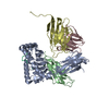
| ||||||||
|---|---|---|---|---|---|---|---|---|---|
| 1 |
| ||||||||
| Unit cell |
| ||||||||
| Details | THE ASYMMETRIC UNIT IS THE BIOLOGICAL COMPLEX, CONSISTING OF CHAINS A,B,P,C,D. |
-Components
-Protein , 2 types, 2 molecules AB
| #1: Protein | Mass: 32007.609 Da / Num. of mol.: 1 / Fragment: Alpha-1,2,3 regions: Residues 26-299 Source method: isolated from a genetically manipulated source Source: (gene. exp.) Mus musculus (house mouse) / Gene: H2-D1 / Plasmid: pET21b / Production host:  Escherichia coli (E. coli) / References: UniProt: P01900 Escherichia coli (E. coli) / References: UniProt: P01900 |
|---|---|
| #2: Protein | Mass: 11791.545 Da / Num. of mol.: 1 / Fragment: Residues 21-119 Source method: isolated from a genetically manipulated source Source: (gene. exp.) Mus musculus (house mouse) / Gene: B2m / Plasmid: pET21b / Production host: Escherichia coli (E. coli) / References: UniProt: Q91XJ8, UniProt: P01887*PLUS |
-T-cell surface glycoprotein CD8 ... , 2 types, 2 molecules CD
| #4: Protein | Mass: 18361.881 Da / Num. of mol.: 1 / Fragment: Ig-like V-type domain: Residues 28-149 Source method: isolated from a genetically manipulated source Source: (gene. exp.) Mus musculus (house mouse) / Gene: Cd8a, Lyt-2, Lyt2 / Plasmid: pET21b / Production host: Escherichia coli (E. coli) / References: UniProt: P01731 |
|---|---|
| #5: Protein | Mass: 16883.535 Da / Num. of mol.: 1 / Fragment: Ig-like V-type domain: Residues 22-141 Source method: isolated from a genetically manipulated source Source: (gene. exp.) Mus musculus (house mouse) / Gene: Cd8b, Cd8b1, Ly-3, Lyt-3, Lyt3 / Plasmid: pET21b / Production host: Escherichia coli (E. coli) / References: UniProt: P10300 |
-Protein/peptide / Non-polymers , 2 types, 38 molecules P
| #3: Protein/peptide | Peptide synthesis Mass: 1075.265 Da / Num. of mol.: 1 / Source method: obtained synthetically / Details: Synthetic peptide |
|---|---|
| #6: Water | ChemComp-HOH / WaterMass: 18.015 Da / Num. of mol.: 37 / Source method: isolated from a natural source / Formula: H2O |
-Experimental details
-Experiment
| Experiment | Method: X-RAY DIFFRACTION / Number of used crystals: 1 |
|---|
- Sample preparation
| Crystal | Density Matthews: 2.34 Å3/Da / Density % sol: 47.4 % |
|---|---|
| Crystal grow | Temperature: 291 K / pH: 7.5 Details: 12% PEG 3000, 0.05M HEPES pH 7.5, VAPOR DIFFUSION, HANGING DROP, temperature 291K |
-Data collection
| Diffraction | Mean temperature: 100 K |
|---|---|
| Diffraction source | Source: SYNCHROTRON / Site: APS  / Beamline: 22-ID / Wavelength: 1 / Beamline: 22-ID / Wavelength: 1 |
| Detector | Type: MAR scanner 300 mm plate / Detector: IMAGE PLATE / Date: Dec 5, 2007 |
| Radiation | Protocol: SINGLE WAVELENGTH / Monochromatic (M) / Laue (L): M / Scattering type: x-ray |
| Radiation wavelength | Wavelength: 1 Å / Relative weight: 1 |
| Reflection | Resolution: 2.6→100 Å / Num. obs: 22842 / % possible obs: 96.1 % / Observed criterion σ(I): 0 / Redundancy: 6.4 % / Biso Wilson estimate: 49.2 Å2 / Rmerge(I) obs: 0.057 / Net I/σ(I): 17.45 |
| Reflection shell | Resolution: 2.6→2.69 Å / Redundancy: 4.2 % / Rmerge(I) obs: 0.432 / Mean I/σ(I) obs: 2.6 / % possible all: 76.9 |
- Processing
| Software |
| ||||||||||||||||||||||||||||||||||||||||||||||||||||||||||||||||||||||||||||||||
|---|---|---|---|---|---|---|---|---|---|---|---|---|---|---|---|---|---|---|---|---|---|---|---|---|---|---|---|---|---|---|---|---|---|---|---|---|---|---|---|---|---|---|---|---|---|---|---|---|---|---|---|---|---|---|---|---|---|---|---|---|---|---|---|---|---|---|---|---|---|---|---|---|---|---|---|---|---|---|---|---|---|
| Refinement | Method to determine structure: MOLECULAR REPLACEMENT Starting model: PDB ENTRIES 1QO3, 2ATP Resolution: 2.6→43.32 Å / Rfactor Rfree error: 0.008 / Occupancy max: 1 / Occupancy min: 1 / Data cutoff high absF: 92554.89 / Isotropic thermal model: RESTRAINED / Cross valid method: THROUGHOUT / σ(F): 0 / Stereochemistry target values: ENGH & HUBER
| ||||||||||||||||||||||||||||||||||||||||||||||||||||||||||||||||||||||||||||||||
| Solvent computation | Solvent model: FLAT / Bsol: 63.66 Å2 / ksol: 0.409086 e/Å3 | ||||||||||||||||||||||||||||||||||||||||||||||||||||||||||||||||||||||||||||||||
| Displacement parameters | Biso mean: 54.7 Å2
| ||||||||||||||||||||||||||||||||||||||||||||||||||||||||||||||||||||||||||||||||
| Refine analyze |
| ||||||||||||||||||||||||||||||||||||||||||||||||||||||||||||||||||||||||||||||||
| Refinement step | Cycle: LAST / Resolution: 2.6→43.32 Å
| ||||||||||||||||||||||||||||||||||||||||||||||||||||||||||||||||||||||||||||||||
| Refine LS restraints |
| ||||||||||||||||||||||||||||||||||||||||||||||||||||||||||||||||||||||||||||||||
| LS refinement shell | Resolution: 2.6→2.76 Å / Rfactor Rfree error: 0.029 / Total num. of bins used: 6
|
