 Movie
Movie Controller

[English] 日本語
 Yorodumi
- PDB-3dah: 2.3 A crystal structure of ribose-phosphate pyrophosphokinase fro... -
+ Open data

- Basic information
| Entry | Database: PDB / ID: 3dah | ||||||
|---|---|---|---|---|---|---|---|
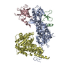
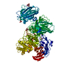
| Title | 2.3 A crystal structure of ribose-phosphate pyrophosphokinase from Burkholderia pseudomallei | ||||||
 Components Components | Ribose-phosphate pyrophosphokinase | ||||||
 Keywords Keywords |  TRANSFERASE / BURKHOLDERIA / PSEUDOMALLEI / RIBOSE / PHOSPHATE / PYROPHOSPHOKINASE / SEATTLE STRUCTURAL GENOMICS CENTER FOR INFECTIOUS DISEASE / SSGCID / Magnesium / Metal binding / Nucleotide biosynthesis TRANSFERASE / BURKHOLDERIA / PSEUDOMALLEI / RIBOSE / PHOSPHATE / PYROPHOSPHOKINASE / SEATTLE STRUCTURAL GENOMICS CENTER FOR INFECTIOUS DISEASE / SSGCID / Magnesium / Metal binding / Nucleotide biosynthesis | ||||||
| Function / homology |  Function and homology informationribose-phosphate diphosphokinase / ribose phosphate diphosphokinase activity / ribonucleoside monophosphate biosynthetic process / nucleotide biosynthetic process / 5-phosphoribose 1-diphosphate biosynthetic process / nucleoside metabolic process / kinase activity / magnesium ion binding / ATP binding / cytoplasm Function and homology informationribose-phosphate diphosphokinase / ribose phosphate diphosphokinase activity / ribonucleoside monophosphate biosynthetic process / nucleotide biosynthetic process / 5-phosphoribose 1-diphosphate biosynthetic process / nucleoside metabolic process / kinase activity / magnesium ion binding / ATP binding / cytoplasmSimilarity search - Function | ||||||
| Biological species |  Burkholderia pseudomallei (bacteria) Burkholderia pseudomallei (bacteria) | ||||||
| Method | X-RAY DIFFRACTION / SYNCHROTRON / MOLECULAR REPLACEMENT / molecular replacement / Resolution: 2.3 Å | ||||||
 Authors Authors | Seattle Structural Genomics Center for Infectious Disease (SSGCID) | ||||||
 Citation Citation | Journal: Plos One / Year: 2013 Title: Combining functional and structural genomics to sample the essential Burkholderia structome. Authors: Baugh, L. / Gallagher, L.A. / Patrapuvich, R. / Clifton, M.C. / Gardberg, A.S. / Edwards, T.E. / Armour, B. / Begley, D.W. / Dieterich, S.H. / Dranow, D.M. / Abendroth, J. / Fairman, J.W. / ...Authors: Baugh, L. / Gallagher, L.A. / Patrapuvich, R. / Clifton, M.C. / Gardberg, A.S. / Edwards, T.E. / Armour, B. / Begley, D.W. / Dieterich, S.H. / Dranow, D.M. / Abendroth, J. / Fairman, J.W. / Fox, D. / Staker, B.L. / Phan, I. / Gillespie, A. / Choi, R. / Nakazawa-Hewitt, S. / Nguyen, M.T. / Napuli, A. / Barrett, L. / Buchko, G.W. / Stacy, R. / Myler, P.J. / Stewart, L.J. / Manoil, C. / Van Voorhis, W.C. | ||||||
| History |
|
- Structure visualization
| Structure viewer | Molecule: MolmilJmol/JSmol |
|---|
- Downloads & links
-Download
| PDBx/mmCIF format | 3dah.cif.gz | 179 KB | Display | PDBx/mmCIF format |
|---|---|---|---|---|
| PDB format | pdb3dah.ent.gz | 142.3 KB | Display | PDB format |
| PDBx/mmJSON format | 3dah.json.gz | Tree view | PDBx/mmJSON format | |
| Others |  Other downloads Other downloads |
-Validation report
| Arichive directory | https://data.pdbj.org/pub/pdb/validation_reports/da/3dahftp://data.pdbj.org/pub/pdb/validation_reports/da/3dah | HTTPS FTP |
|---|
-Related structure data
| Related structure data | 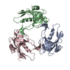 3d63C 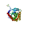 3eizC 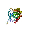 3ej2C 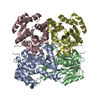 3ek2C  3ezoC  3f0fC  3ftpC  3gk0C 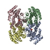 3gk3C 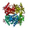 3gvfC 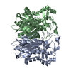 3gwaC 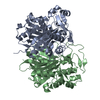 3gweC  3imlC  3sz8C  3t4cC  3tmlC 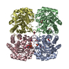 3tmqC 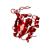 3txyC 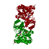 3u7jC  3ue9C 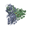 3uk1C  3uk2C  3undC  3uptC  3urrC  3uw1C 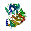 3uw2C 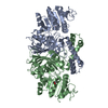 3uw3C 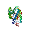 3v2iC 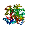 3v7nC 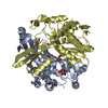 3v8hC 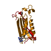 3v9oC 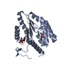 3v9pC 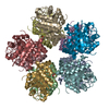 3vavC 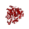 4ddoC 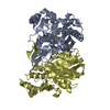 4dfeC  4dheC 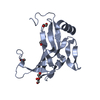 4dhkC 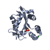 4dutC  4dz4C  4e4tC  4efiC  4eg0C 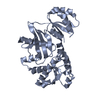 4egjC 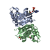 4ek2C 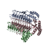 4eqyC 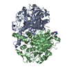 4ewgC  4exqC  4f2gC  4f32C  4f3nC  4f3yC  4f4hC  4f7dC 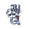 4fk8C  4fryC  4g1kC 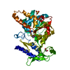 4g67C 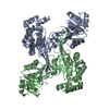 4ghkC  4h3yC  4h3zC  4h4gC  1dkuS S: Starting model for refinement C: citing same article ( |
|---|---|
| Similar structure data | |
| Other databases |


-Links
 PDBj
PDBj


- Assembly
| Deposited unit | 
| ||||||||
|---|---|---|---|---|---|---|---|---|---|
| 1 |
| ||||||||
| 2 | 
| ||||||||
| Unit cell |
|
-Components
| #1: Protein | Mass: 34335.734 Da / Num. of mol.: 3 Source method: isolated from a genetically manipulated source Source: (gene. exp.) Burkholderia pseudomallei (bacteria) / Strain: 1710b / Gene: prsA, BURPS1710b_0753 / Plasmid: Ava0421 / Production host: Escherichia coli (E. coli) / Strain (production host): BL21(DE3)References: UniProt: Q3JW86, UniProt: Q63XL8*PLUS, ribose-phosphate diphosphokinase#2: Chemical | ChemComp-PO4 / Phosphate  Mass: 94.971 Da / Num. of mol.: 7 / Source method: obtained synthetically / Formula: PO4 Mass: 94.971 Da / Num. of mol.: 7 / Source method: obtained synthetically / Formula: PO4#3: Chemical | Adenosine monophosphate  Mass: 347.221 Da / Num. of mol.: 3 / Source method: obtained synthetically / Formula: C10H14N5O7P / Comment: AMP*YM Mass: 347.221 Da / Num. of mol.: 3 / Source method: obtained synthetically / Formula: C10H14N5O7P / Comment: AMP*YM#4: Water | ChemComp-HOH / | Water Mass: 18.015 Da / Num. of mol.: 138 / Source method: isolated from a natural source / Formula: H2O Mass: 18.015 Da / Num. of mol.: 138 / Source method: isolated from a natural source / Formula: H2O |
|---|
-Experimental details
-Experiment
| Experiment | Method: X-RAY DIFFRACTION / Number of used crystals: 1 |
|---|
- Sample preparation
| Crystal | Density Matthews: 2.3 Å3/Da / Density % sol: 46.55 % |
|---|---|
| Crystal grow | Temperature: 289 K / Method: vapor diffusion / pH: 5.9 Details: 20% PEG 3350, 0.2M Magnesium formate, pH 5.9, VAPOR DIFFUSION, temperature 289K |
-Data collection
| Diffraction | Mean temperature: 100 K | |||||||||||||||||||||||||||||||||||||||||||||||||||||||||||||||||||||||||||||
|---|---|---|---|---|---|---|---|---|---|---|---|---|---|---|---|---|---|---|---|---|---|---|---|---|---|---|---|---|---|---|---|---|---|---|---|---|---|---|---|---|---|---|---|---|---|---|---|---|---|---|---|---|---|---|---|---|---|---|---|---|---|---|---|---|---|---|---|---|---|---|---|---|---|---|---|---|---|---|
| Diffraction source | Source: SYNCHROTRON / Site: APS  / Beamline: 23-ID-D / Wavelength: 1 Å / Beamline: 23-ID-D / Wavelength: 1 Å | |||||||||||||||||||||||||||||||||||||||||||||||||||||||||||||||||||||||||||||
| Detector | Type: MARMOSAIC 300 mm CCD / Detector: CCD / Date: Apr 2, 2008 / Details: ADJUSTABLE FOCUSING MIRRORS | |||||||||||||||||||||||||||||||||||||||||||||||||||||||||||||||||||||||||||||
| Radiation | Monochromator: Double crystal / Protocol: SINGLE WAVELENGTH / Monochromatic (M) / Laue (L): M / Scattering type: x-ray | |||||||||||||||||||||||||||||||||||||||||||||||||||||||||||||||||||||||||||||
| Radiation wavelength | Wavelength: 1 Å / Relative weight: 1 | |||||||||||||||||||||||||||||||||||||||||||||||||||||||||||||||||||||||||||||
| Reflection | Resolution: 2.3→50 Å / Num. obs: 41722 / % possible obs: 98 % / Redundancy: 6 % / Rmerge(I) obs: 0.09 / Χ2: 0.926 / Net I/σ(I): 7.9 | |||||||||||||||||||||||||||||||||||||||||||||||||||||||||||||||||||||||||||||
| Reflection shell |
|
-Phasing
| Phasing | Method: molecular replacement | |||||||||
|---|---|---|---|---|---|---|---|---|---|---|
| Phasing MR |
|
- Processing
| Software |
| ||||||||||||||||||||||||||||||||||||||||||||||||||||||||||||||||||||||||||||||||||||||||||
|---|---|---|---|---|---|---|---|---|---|---|---|---|---|---|---|---|---|---|---|---|---|---|---|---|---|---|---|---|---|---|---|---|---|---|---|---|---|---|---|---|---|---|---|---|---|---|---|---|---|---|---|---|---|---|---|---|---|---|---|---|---|---|---|---|---|---|---|---|---|---|---|---|---|---|---|---|---|---|---|---|---|---|---|---|---|---|---|---|---|---|---|
| Refinement | Method to determine structure: MOLECULAR REPLACEMENT Starting model: PDB entry 1DKU Resolution: 2.3→46.93 Å / Cor.coef. Fo:Fc: 0.951 / Cor.coef. Fo:Fc free: 0.927 / SU B: 8.489 / SU ML: 0.202 / Cross valid method: THROUGHOUT / σ(F): 0 / ESU R: 0.367 / ESU R Free: 0.251 / Stereochemistry target values: MAXIMUM LIKELIHOOD
| ||||||||||||||||||||||||||||||||||||||||||||||||||||||||||||||||||||||||||||||||||||||||||
| Solvent computation | Ion probe radii: 0.8 Å / Shrinkage radii: 0.8 Å / VDW probe radii: 1.2 Å / Solvent model: MASK | ||||||||||||||||||||||||||||||||||||||||||||||||||||||||||||||||||||||||||||||||||||||||||
| Displacement parameters | Biso mean: 48.513 Å2
| ||||||||||||||||||||||||||||||||||||||||||||||||||||||||||||||||||||||||||||||||||||||||||
| Refinement step | Cycle: LAST / Resolution: 2.3→46.93 Å
| ||||||||||||||||||||||||||||||||||||||||||||||||||||||||||||||||||||||||||||||||||||||||||
| Refine LS restraints |
| ||||||||||||||||||||||||||||||||||||||||||||||||||||||||||||||||||||||||||||||||||||||||||
| LS refinement shell | Resolution: 2.3→2.361 Å / Total num. of bins used: 20
|
