 Movie
Movie Controller

[English] 日本語
 Yorodumi
- PDB-3d7a: Crystal structure of DUF54 family protein PH1010 from hyperthermo... -
+ Open data

- Basic information
| Entry | Database: PDB / ID: 3d7a | ||||||
|---|---|---|---|---|---|---|---|
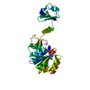
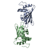
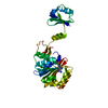
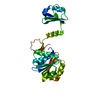
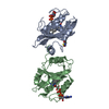
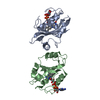
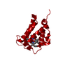
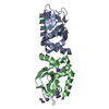
| Title | Crystal structure of DUF54 family protein PH1010 from hyperthermophilic archaea Pyrococcus horikoshii OT3 | ||||||
 Components Components | UPF0201 protein PH1010 | ||||||
 Keywords Keywords | UNKNOWN FUNCTION / DUF54 family | ||||||
| Function / homology | Uncharacterised protein family UPF0201 /  RNA binding / 50s Ribosomal Protein L5; Chain: A, / Ribosomal protein L5 / Ribosomal protein L5 domain superfamily / 2-Layer Sandwich / Alpha Beta / UPF0201 protein PH1010 RNA binding / 50s Ribosomal Protein L5; Chain: A, / Ribosomal protein L5 / Ribosomal protein L5 domain superfamily / 2-Layer Sandwich / Alpha Beta / UPF0201 protein PH1010 Function and homology information Function and homology information | ||||||
| Biological species |   Pyrococcus horikoshii (archaea) Pyrococcus horikoshii (archaea) | ||||||
| Method | X-RAY DIFFRACTION / SYNCHROTRON / Resolution: 1.9 Å | ||||||
 Authors Authors | Shirokane, M. / Miyazono, K.I. / Sawano, Y. / Tanokura, M. | ||||||
 Citation Citation | Journal: Proteins / Year: 2008 Title: Crystal structure of the DUF54 family protein PH1010 from hyperthermophilic archaea Pyrococcus horikoshii OT3. Authors: Miyazono, K.I. / Shirokane, M. / Sawano, Y. / Tanokura, M. | ||||||
| History |
|
- Structure visualization
| Structure viewer | Molecule: MolmilJmol/JSmol |
|---|
- Downloads & links
-Download
| PDBx/mmCIF format | 3d7a.cif.gz | 69.8 KB | Display | PDBx/mmCIF format |
|---|---|---|---|---|
| PDB format | pdb3d7a.ent.gz | 52.3 KB | Display | PDB format |
| PDBx/mmJSON format | 3d7a.json.gz | Tree view | PDBx/mmJSON format | |
| Others |  Other downloads Other downloads |
-Validation report
| Arichive directory | https://data.pdbj.org/pub/pdb/validation_reports/d7/3d7aftp://data.pdbj.org/pub/pdb/validation_reports/d7/3d7a | HTTPS FTP |
|---|
-Related structure data
| Similar structure data |
|---|


-Links
 PDBj
PDBj- Assembly
| Deposited unit | 
| ||||||||
|---|---|---|---|---|---|---|---|---|---|
| 1 |
| ||||||||
| Unit cell |
|
-Components
| #1: Protein | Mass: 16077.828 Da / Num. of mol.: 2 Source method: isolated from a genetically manipulated source Source: (gene. exp.) Pyrococcus horikoshii (archaea) / Strain: OT3 / Gene: PH1010 / Production host:  Escherichia coli (E. coli) / References: UniProt: O58738 Escherichia coli (E. coli) / References: UniProt: O58738#2: Water | ChemComp-HOH / | Water Mass: 18.015 Da / Num. of mol.: 176 / Source method: isolated from a natural source / Formula: H2O Mass: 18.015 Da / Num. of mol.: 176 / Source method: isolated from a natural source / Formula: H2O |
|---|
-Experimental details
-Experiment
| Experiment | Method: X-RAY DIFFRACTION / Number of used crystals: 1 |
|---|
- Sample preparation
| Crystal | Density Matthews: 2.4 Å3/Da / Density % sol: 48.75 % / Mosaicity: 0.259 ° |
|---|
-Data collection
| Diffraction source | Source: SYNCHROTRON / Site: Photon Factory  / Beamline: BL-5A / Wavelength: 1 Å / Beamline: BL-5A / Wavelength: 1 Å | ||||||||||||||||||||||||||||||||||||||||||||||||||||||||||||||||||
|---|---|---|---|---|---|---|---|---|---|---|---|---|---|---|---|---|---|---|---|---|---|---|---|---|---|---|---|---|---|---|---|---|---|---|---|---|---|---|---|---|---|---|---|---|---|---|---|---|---|---|---|---|---|---|---|---|---|---|---|---|---|---|---|---|---|---|---|
| Radiation | Protocol: SINGLE WAVELENGTH / Monochromatic (M) / Laue (L): M / Scattering type: x-ray | ||||||||||||||||||||||||||||||||||||||||||||||||||||||||||||||||||
| Radiation wavelength | Wavelength: 1 Å / Relative weight: 1 | ||||||||||||||||||||||||||||||||||||||||||||||||||||||||||||||||||
| Reflection | Resolution: 1.9→30 Å / Num. obs: 25090 / % possible obs: 99.7 % / Redundancy: 6.9 % / Rmerge(I) obs: 0.061 / Χ2: 2.068 / Net I/σ(I): 16.5 | ||||||||||||||||||||||||||||||||||||||||||||||||||||||||||||||||||
| Reflection shell |
|
- Processing
| Software |
| ||||||||||||||||||||||||||||||||||||||||||||||||||||||||||||||||||||||||||||||||||||||||||||||||||||||||||||||||||||||||||||||||||||||||||||||||||||||||||||||||||||||||||
|---|---|---|---|---|---|---|---|---|---|---|---|---|---|---|---|---|---|---|---|---|---|---|---|---|---|---|---|---|---|---|---|---|---|---|---|---|---|---|---|---|---|---|---|---|---|---|---|---|---|---|---|---|---|---|---|---|---|---|---|---|---|---|---|---|---|---|---|---|---|---|---|---|---|---|---|---|---|---|---|---|---|---|---|---|---|---|---|---|---|---|---|---|---|---|---|---|---|---|---|---|---|---|---|---|---|---|---|---|---|---|---|---|---|---|---|---|---|---|---|---|---|---|---|---|---|---|---|---|---|---|---|---|---|---|---|---|---|---|---|---|---|---|---|---|---|---|---|---|---|---|---|---|---|---|---|---|---|---|---|---|---|---|---|---|---|---|---|---|---|---|---|
| Refinement | Resolution: 1.9→30 Å / Cor.coef. Fo:Fc: 0.938 / Cor.coef. Fo:Fc free: 0.924 / SU B: 4.498 / SU ML: 0.133 / Cross valid method: THROUGHOUT / σ(F): 0 / ESU R: 0.189 / ESU R Free: 0.172 / Stereochemistry target values: MAXIMUM LIKELIHOOD
| ||||||||||||||||||||||||||||||||||||||||||||||||||||||||||||||||||||||||||||||||||||||||||||||||||||||||||||||||||||||||||||||||||||||||||||||||||||||||||||||||||||||||||
| Solvent computation | Ion probe radii: 0.8 Å / Shrinkage radii: 0.8 Å / VDW probe radii: 1.4 Å / Solvent model: MASK | ||||||||||||||||||||||||||||||||||||||||||||||||||||||||||||||||||||||||||||||||||||||||||||||||||||||||||||||||||||||||||||||||||||||||||||||||||||||||||||||||||||||||||
| Displacement parameters | Biso mean: 33.019 Å2
| ||||||||||||||||||||||||||||||||||||||||||||||||||||||||||||||||||||||||||||||||||||||||||||||||||||||||||||||||||||||||||||||||||||||||||||||||||||||||||||||||||||||||||
| Refinement step | Cycle: LAST / Resolution: 1.9→30 Å
| ||||||||||||||||||||||||||||||||||||||||||||||||||||||||||||||||||||||||||||||||||||||||||||||||||||||||||||||||||||||||||||||||||||||||||||||||||||||||||||||||||||||||||
| Refine LS restraints |
| ||||||||||||||||||||||||||||||||||||||||||||||||||||||||||||||||||||||||||||||||||||||||||||||||||||||||||||||||||||||||||||||||||||||||||||||||||||||||||||||||||||||||||
| LS refinement shell | Resolution: 1.9→1.949 Å / Total num. of bins used: 20
|
