 Movie
Movie Controller


+ Open data

- Basic information
| Entry | Database: PDB / ID: 4uzv | ||||||
|---|---|---|---|---|---|---|---|
| Title | Structure of a triple mutant of ASV-TfTrHb | ||||||
 Components Components | HEMOGLOBIN | ||||||
 Keywords Keywords | OXIDOREDUCTASE / BACTERIAL HEMOGLOBINS / HEME LIGAND-BINDING PROPERTIES / THERMOSTABLEPROTEINS | ||||||
| Function / homology | Globins / Globin-like / Orthogonal Bundle / Mainly Alpha / ACETATE ION / PROTOPORPHYRIN IX CONTAINING FE / :  Function and homology information Function and homology information | ||||||
| Biological species |   THERMOBIFIDA FUSCA TM51 (bacteria) THERMOBIFIDA FUSCA TM51 (bacteria) | ||||||
| Method | X-RAY DIFFRACTION / SYNCHROTRON / MOLECULAR REPLACEMENT / Resolution: 3.4 Å | ||||||
 Authors Authors | Baiocco, P. / Bonamore, A. / Sciamanna, N. / Ilari, A. / Boechi, L. / Boffi, A. / Smulevich, G. / Feis, A. | ||||||
 Citation Citation | Journal: J.Phys.Chem.B / Year: 2014 Title: Role of Local Structure and Dynamics of Small Ligand Migration in Proteins: A Study of a Mutated Truncated Hemoprotein from Thermobifida Fusca by Time Resolved Mir Spectroscopy. Authors: Patrizi, B. / Lapini, A. / Di Donato, M. / Marcelli, A. / Lima, M. / Righini, R. / Foggi, P. / Baiocco, P. / Bonamore, A. / Boffi, A. | ||||||
| History |
|
- Structure visualization
| Structure viewer | Molecule: MolmilJmol/JSmol |
|---|
- Downloads & links
-Download
| PDBx/mmCIF format | 4uzv.cif.gz | 38.4 KB | Display | PDBx/mmCIF format |
|---|---|---|---|---|
| PDB format | pdb4uzv.ent.gz | 25.9 KB | Display | PDB format |
| PDBx/mmJSON format | 4uzv.json.gz | Tree view | PDBx/mmJSON format | |
| Others |  Other downloads Other downloads |
-Validation report
| Arichive directory | https://data.pdbj.org/pub/pdb/validation_reports/uz/4uzvftp://data.pdbj.org/pub/pdb/validation_reports/uz/4uzv | HTTPS FTP |
|---|
-Related structure data
| Related structure data |  2bmmS S: Starting model for refinement |
|---|---|
| Similar structure data |


-Links
 PDBj
PDBj- Assembly
| Deposited unit | 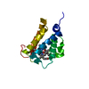
| ||||||||
|---|---|---|---|---|---|---|---|---|---|
| 1 | 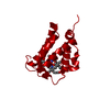
| ||||||||
| Unit cell |
|
-Components
| #1: Protein | Mass: 18970.402 Da / Num. of mol.: 1 / Fragment: UNP RESIDUES 1-164 / Mutation: YES Source method: isolated from a genetically manipulated source Source: (gene. exp.) THERMOBIFIDA FUSCA TM51 (bacteria) / Production host: ESCHERICHIA COLI (E. coli) / Strain (production host): BL21(DE3) / References: UniProt: R9F4S3 |
|---|---|
| #2: Chemical | ChemComp-HEM / Heme B  Mass: 616.487 Da / Num. of mol.: 1 / Source method: obtained synthetically / Formula: C34H32FeN4O4 Mass: 616.487 Da / Num. of mol.: 1 / Source method: obtained synthetically / Formula: C34H32FeN4O4 |
| #3: Chemical | ChemComp-ACT / Acetate  Mass: 59.044 Da / Num. of mol.: 1 / Source method: obtained synthetically / Formula: C2H3O2 Mass: 59.044 Da / Num. of mol.: 1 / Source method: obtained synthetically / Formula: C2H3O2 |
-Experimental details
-Experiment
| Experiment | Method: X-RAY DIFFRACTION |
|---|
- Sample preparation
| Crystal | Density Matthews: 3.93 Å3/Da / Density % sol: 68.7 % / Description: NONE |
|---|
-Data collection
| Diffraction | Mean temperature: 100 K |
|---|---|
| Diffraction source | Source: SYNCHROTRON / Site: BESSY  / Beamline: 14.1 / Wavelength: 0.893 / Beamline: 14.1 / Wavelength: 0.893 |
| Radiation | Protocol: SINGLE WAVELENGTH / Monochromatic (M) / Laue (L): M / Scattering type: x-ray |
| Radiation wavelength | Wavelength: 0.893 Å / Relative weight: 1 |
| Reflection | Resolution: 3.4→50 Å / Num. obs: 4156 / % possible obs: 99.8 % / Observed criterion σ(I): 2 / Redundancy: 14.2 % / Rmerge(I) obs: 0.13 / Net I/σ(I): 25 |
| Reflection shell | Resolution: 3.4→3.52 Å / Redundancy: 14.4 % / Rmerge(I) obs: 0.32 / Mean I/σ(I) obs: 5.8 / % possible all: 99.7 |
- Processing
| Software |
| ||||||||||||||||||||||||||||||||||||||||||||||||||||||||||||||||||||||||||||||||||||||||||||||||||||||||||||||||||||||||||||||||||||||||||||||||||||||||||||||||||||||||||||||||||||||
|---|---|---|---|---|---|---|---|---|---|---|---|---|---|---|---|---|---|---|---|---|---|---|---|---|---|---|---|---|---|---|---|---|---|---|---|---|---|---|---|---|---|---|---|---|---|---|---|---|---|---|---|---|---|---|---|---|---|---|---|---|---|---|---|---|---|---|---|---|---|---|---|---|---|---|---|---|---|---|---|---|---|---|---|---|---|---|---|---|---|---|---|---|---|---|---|---|---|---|---|---|---|---|---|---|---|---|---|---|---|---|---|---|---|---|---|---|---|---|---|---|---|---|---|---|---|---|---|---|---|---|---|---|---|---|---|---|---|---|---|---|---|---|---|---|---|---|---|---|---|---|---|---|---|---|---|---|---|---|---|---|---|---|---|---|---|---|---|---|---|---|---|---|---|---|---|---|---|---|---|---|---|---|---|
| Refinement | Method to determine structure: MOLECULAR REPLACEMENT Starting model: PDB ENTRY 2BMM Resolution: 3.4→50 Å / Cor.coef. Fo:Fc: 0.88 / Cor.coef. Fo:Fc free: 0.872 / SU B: 45.264 / SU ML: 0.683 / Cross valid method: THROUGHOUT / ESU R: 1.412 / ESU R Free: 0.642 / Stereochemistry target values: MAXIMUM LIKELIHOOD / Details: HYDROGENS HAVE BEEN ADDED IN THE RIDING POSITIONS.
| ||||||||||||||||||||||||||||||||||||||||||||||||||||||||||||||||||||||||||||||||||||||||||||||||||||||||||||||||||||||||||||||||||||||||||||||||||||||||||||||||||||||||||||||||||||||
| Solvent computation | Ion probe radii: 0.8 Å / Shrinkage radii: 0.8 Å / VDW probe radii: 1.4 Å / Solvent model: MASK | ||||||||||||||||||||||||||||||||||||||||||||||||||||||||||||||||||||||||||||||||||||||||||||||||||||||||||||||||||||||||||||||||||||||||||||||||||||||||||||||||||||||||||||||||||||||
| Displacement parameters | Biso mean: 87.051 Å2
| ||||||||||||||||||||||||||||||||||||||||||||||||||||||||||||||||||||||||||||||||||||||||||||||||||||||||||||||||||||||||||||||||||||||||||||||||||||||||||||||||||||||||||||||||||||||
| Refinement step | Cycle: LAST / Resolution: 3.4→50 Å
| ||||||||||||||||||||||||||||||||||||||||||||||||||||||||||||||||||||||||||||||||||||||||||||||||||||||||||||||||||||||||||||||||||||||||||||||||||||||||||||||||||||||||||||||||||||||
| Refine LS restraints |
|