Type: MAR CCD 165 mm / Detector: CCD / Date: Apr 4, 2008
Radiation
Protocol: SINGLE WAVELENGTH / Monochromatic (M) / Laue (L): M / Scattering type: x-ray
Radiation wavelength
Wavelength: 0.9798 Å / Relative weight: 1
Reflection
Redundancy: 3.5 % / Av σ(I) over netI: 7.8 / Number: 713527 / Rmerge(I) obs: 0.076 / Χ2: 1.37 / D res high: 1.41 Å / D res low: 50 Å / Num. obs: 206130 / % possible obs: 96.6
Diffraction reflection shell
Highest resolution (Å)
Lowest resolution (Å)
% possible obs (%)
ID
Rmerge(I) obs
Chi squared
Redundancy
3.04
50
99.4
1
0.049
3.052
3.9
2.41
3.04
100
1
0.065
1.903
3.9
2.11
2.41
100
1
0.079
1.401
3.9
1.91
2.11
100
1
0.109
1.063
3.9
1.78
1.91
100
1
0.168
0.846
3.8
1.67
1.78
100
1
0.248
0.719
3.8
1.59
1.67
99.7
1
0.31
0.642
3.5
1.52
1.59
98.4
1
0.39
0.636
3.1
1.46
1.52
96.6
1
0.547
0.798
2.6
1.41
1.46
72.2
1
4.345
1.8
Reflection
Resolution: 1.41→50 Å / Num. obs: 206130 / % possible obs: 96.6 % / Redundancy: 3.5 % / Rmerge(I) obs: 0.076 / Χ2: 1.368 / Net I/σ(I): 7.8
Reflection shell
Resolution (Å)
Redundancy (%)
Num. unique all
Χ2
Diffraction-ID
% possible all
Rmerge(I) obs
1.41-1.46
1.8
15467
4.345
1
72.2
1.46-1.52
2.6
20595
0.798
1
96.6
0.547
1.52-1.59
3.1
20970
0.636
1
98.4
0.39
1.59-1.67
3.5
21211
0.642
1
99.7
0.31
1.67-1.78
3.8
21334
0.719
1
100
0.248
1.78-1.91
3.8
21301
0.846
1
100
0.168
1.91-2.11
3.9
21349
1.063
1
100
0.109
2.11-2.41
3.9
21373
1.401
1
100
0.079
2.41-3.04
3.9
21323
1.903
1
100
0.065
3.04-50
3.9
21207
3.052
1
99.4
0.049
-
Phasing
Phasing
Method: SAD
-
Processing
Software
Name
Version
Classification
NB
DENZO
datareduction
SCALEPACK
datascaling
SHELX
phasing
REFMAC
refinement
PDB_EXTRACT
3.005
dataextraction
MAR345
CCD
datacollection
HKL-2000
datareduction
SHELXD
phasing
Refinement
Method to determine structure: SAD / Resolution: 1.41→9.99 Å / Cor.coef. Fo:Fc: 0.964 / Cor.coef. Fo:Fc free: 0.954 / SU B: 2.713 / SU ML: 0.048 / TLS residual ADP flag: LIKELY RESIDUAL / Cross valid method: THROUGHOUT / σ(F): 0 / ESU R: 0.066 / ESU R Free: 0.067 / Stereochemistry target values: MAXIMUM LIKELIHOOD / Details: The Friedel pairs were used in phasing.
Rfactor
Num. reflection
% reflection
Selection details
Rfree
0.207
5140
5 %
RANDOM
Rwork
0.182
-
-
-
obs
0.183
102645
94.93 %
-
Solvent computation
Ion probe radii: 0.8 Å / Shrinkage radii: 0.8 Å / VDW probe radii: 1.2 Å / Solvent model: BABINET MODEL WITH MASK
Displacement parameters
Biso mean: 17.288 Å2
Baniso -1
Baniso -2
Baniso -3
1-
0 Å2
0 Å2
0 Å2
2-
-
0 Å2
0 Å2
3-
-
-
-0.01 Å2
Refinement step
Cycle: LAST / Resolution: 1.41→9.99 Å
Protein
Nucleic acid
Ligand
Solvent
Total
Num. atoms
4254
0
22
489
4765
Refine LS restraints
Refine-ID
Type
Dev ideal
Dev ideal target
Number
X-RAY DIFFRACTION
r_bond_refined_d
0.014
0.022
4430
X-RAY DIFFRACTION
r_angle_refined_deg
1.51
1.971
6001
X-RAY DIFFRACTION
r_dihedral_angle_1_deg
6.132
5
549
X-RAY DIFFRACTION
r_dihedral_angle_2_deg
36.819
25.535
215
X-RAY DIFFRACTION
r_dihedral_angle_3_deg
12.906
15
808
X-RAY DIFFRACTION
r_dihedral_angle_4_deg
11.265
15
18
X-RAY DIFFRACTION
r_chiral_restr
0.11
0.2
685
X-RAY DIFFRACTION
r_gen_planes_refined
0.007
0.021
3326
X-RAY DIFFRACTION
r_mcbond_it
1.347
1.5
2706
X-RAY DIFFRACTION
r_mcangle_it
3.736
20
4405
X-RAY DIFFRACTION
r_scbond_it
6.33
20
1724
X-RAY DIFFRACTION
r_scangle_it
4.834
4.5
1596
LS refinement shell
Resolution: 1.41→1.446 Å / Total num. of bins used: 20
Rfactor
Num. reflection
% reflection
Rfree
0.573
200
-
Rwork
0.524
3855
-
all
-
4055
-
obs
-
-
51.6 %
Refinement TLS params.
Method: refined / Refine-ID: X-RAY DIFFRACTION
ID
L11 (°2)
L12 (°2)
L13 (°2)
L22 (°2)
L23 (°2)
L33 (°2)
S11 (Å °)
S12 (Å °)
S13 (Å °)
S21 (Å °)
S22 (Å °)
S23 (Å °)
S31 (Å °)
S32 (Å °)
S33 (Å °)
T11 (Å2)
T12 (Å2)
T13 (Å2)
T22 (Å2)
T23 (Å2)
T33 (Å2)
Origin x (Å)
Origin y (Å)
Origin z (Å)
1
0.2299
-0.0611
0.0883
0.1199
0.0275
0.1959
-0.0109
0.0215
-0.0131
0.0151
0.0014
0.0132
0.0088
0
0.0095
-0.0067
-0.0024
0.0038
-0.0118
-0.0021
-0.0074
26.3922
29.4609
4.7689
2
0.3404
0.0274
-0.0557
0.1249
0.0451
0.1278
-0.0099
-0.0206
0.0034
-0.0243
0.0018
0.004
-0.0181
-0.0098
0.008
-0.0033
0.0015
-0.0021
-0.0113
-0.001
-0.0122
20.0503
41.923
29.0128
3
0.0436
-0.0282
0.0176
0.0182
-0.0114
0.0071
0.0384
0.054
0.0005
0.0089
-0.0562
-0.0185
0.0152
-0.2287
0.0178
0.0023
-0.003
0.0008
0.0096
-0.0091
0.0027
20.8677
35.6885
16.2578
Refinement TLS group
ID
Refine-ID
Refine TLS-ID
Auth asym-ID
Auth seq-ID
1
X-RAY DIFFRACTION
1
A
60 - 325
2
X-RAY DIFFRACTION
2
B
60 - 325
3
X-RAY DIFFRACTION
3
A
401 - 402
4
X-RAY DIFFRACTION
3
B
401 - 402
+
About Yorodumi
-
News
-
Feb 9, 2022. New format data for meta-information of EMDB entries
New format data for meta-information of EMDB entries
Version 3 of the EMDB header file is now the official format.
The previous official version 1.9 will be removed from the archive.
In the structure databanks used in Yorodumi, some data are registered as the other names, "COVID-19 virus" and "2019-nCoV". Here are the details of the virus and the list of structure data.
Jan 31, 2019. EMDB accession codes are about to change! (news from PDBe EMDB page)
EMDB accession codes are about to change! (news from PDBe EMDB page)
The allocation of 4 digits for EMDB accession codes will soon come to an end. Whilst these codes will remain in use, new EMDB accession codes will include an additional digit and will expand incrementally as the available range of codes is exhausted. The current 4-digit format prefixed with “EMD-” (i.e. EMD-XXXX) will advance to a 5-digit format (i.e. EMD-XXXXX), and so on. It is currently estimated that the 4-digit codes will be depleted around Spring 2019, at which point the 5-digit format will come into force.
The EM Navigator/Yorodumi systems omit the EMD- prefix.
Related info.:Q: What is EMD? / ID/Accession-code notation in Yorodumi/EM Navigator
Yorodumi is a browser for structure data from EMDB, PDB, SASBDB, etc.
This page is also the successor to EM Navigator detail page, and also detail information page/front-end page for Omokage search.
The word "yorodu" (or yorozu) is an old Japanese word meaning "ten thousand". "mi" (miru) is to see.
Related info.:EMDB / PDB / SASBDB / Comparison of 3 databanks / Yorodumi Search / Aug 31, 2016. New EM Navigator & Yorodumi / Yorodumi Papers / Jmol/JSmol / Function and homology information / Changes in new EM Navigator and Yorodumi
 Movie
Movie Controller

 Yorodumi
 Open data
 Basic information
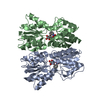
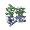
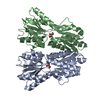
 Components
 Keywords
 TRANSCRIPTION REGULATOR /
  Function and homology information
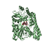
 Alkaliphilus metalliredigens (bacteria)
 Authors
 Citation
 Structure visualization
 Downloads & links
 Other downloads
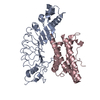


 PDBj
PDBj


 Assembly
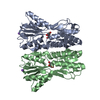

 Type: D-saccharide, beta linking / Mass: 150.130 Da / Num. of mol.: 2 / Source method: obtained synthetically / Formula: C5H10O5
 Mass: 22.990 Da / Num. of mol.: 2 / Source method: obtained synthetically / Formula: Na
 Mass: 18.015 Da / Num. of mol.: 489 / Source method: isolated from a natural source / Formula: H2O
 Sample preparation
 / Beamline: 31-ID / Wavelength: 0.9798 Å
 Processing