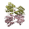
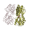
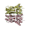
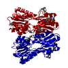
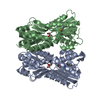
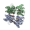
- PDB-3c3k: Crystal structure of an uncharacterized protein from Actinobacill... -
+
Open data
ID or keywords:
Loading...
-
Basic information
Entry
Database: PDB / ID: 3c3k
Title
Crystal structure of an uncharacterized protein from Actinobacillus succinogenes
Components
Alanine racemase
Keywords
ISOMERASE / STRUCTURAL GENOMICS / PROTEIN STRUCTURE INITIATIVE / New York SGX Research Center for Structural Genomics / NYSGXRC / DNA-binding / Transcription / Transcription regulation / PSI-2
Function / homology
Function and homology information
alanine racemase / alanine racemase activity / regulation of DNA-templated transcription / DNA binding Similarity search - Function
Protocol: SINGLE WAVELENGTH / Monochromatic (M) / Laue (L): M / Scattering type: x-ray
Radiation wavelength
Wavelength: 0.979 Å / Relative weight: 1
Reflection
Redundancy: 3 % / Av σ(I) over netI: 8.4 / Number: 165629 / Rmerge(I) obs: 0.088 / Χ2: 1.28 / D res high: 1.99 Å / D res low: 50 Å / Num. obs: 55962 / % possible obs: 86.4
Diffraction reflection shell
Highest resolution (Å)
Lowest resolution (Å)
% possible obs (%)
ID
Rmerge(I) obs
Chi squared
Redundancy
4.29
50
99.7
1
0.048
1.841
3.6
3.4
4.29
99.7
1
0.068
2.189
3.4
2.97
3.4
99.7
1
0.086
1.37
3.3
2.7
2.97
99.9
1
0.12
1.063
3.3
2.51
2.7
99.6
1
0.165
0.886
3.2
2.36
2.51
98.3
1
0.194
0.851
2.9
2.24
2.36
92.8
1
0.22
0.807
2.6
2.14
2.24
78.5
1
0.232
0.761
2.2
2.06
2.14
57.5
1
0.241
0.792
2
1.99
2.06
38.4
1
0.245
0.806
1.8
Reflection
Resolution: 1.99→50 Å / Num. obs: 55962 / % possible obs: 86.4 % / Redundancy: 3 % / Rmerge(I) obs: 0.088 / Χ2: 1.283 / Net I/σ(I): 8.4
Method to determine structure: SAD / Resolution: 1.99→30.47 Å / Cor.coef. Fo:Fc: 0.95 / Cor.coef. Fo:Fc free: 0.925 / SU B: 4.527 / SU ML: 0.129 / Cross valid method: THROUGHOUT / σ(F): 0 / ESU R: 0.219 / ESU R Free: 0.18 / Stereochemistry target values: MAXIMUM LIKELIHOOD / Details: The Friedel pairs were used in phasing
Rfactor
Num. reflection
% reflection
Selection details
Rfree
0.231
1596
5.1 %
RANDOM
Rwork
0.18
-
-
-
obs
0.183
31315
100 %
-
Solvent computation
Ion probe radii: 0.8 Å / Shrinkage radii: 0.8 Å / VDW probe radii: 1.2 Å / Solvent model: BABINET MODEL WITH MASK
Displacement parameters
Biso mean: 19.901 Å2
Baniso -1
Baniso -2
Baniso -3
1-
0.02 Å2
0 Å2
0 Å2
2-
-
-0.03 Å2
0 Å2
3-
-
-
0.01 Å2
Refinement step
Cycle: LAST / Resolution: 1.99→30.47 Å
Protein
Nucleic acid
Ligand
Solvent
Total
Num. atoms
4120
0
13
253
4386
Refine LS restraints
Refine-ID
Type
Dev ideal
Dev ideal target
Number
X-RAY DIFFRACTION
r_bond_refined_d
0.022
0.022
4197
X-RAY DIFFRACTION
r_angle_refined_deg
1.994
1.974
5690
X-RAY DIFFRACTION
r_dihedral_angle_1_deg
6.395
5
536
X-RAY DIFFRACTION
r_dihedral_angle_2_deg
38.785
24.94
166
X-RAY DIFFRACTION
r_dihedral_angle_3_deg
16.446
15
736
X-RAY DIFFRACTION
r_dihedral_angle_4_deg
17.973
15
18
X-RAY DIFFRACTION
r_chiral_restr
0.156
0.2
680
X-RAY DIFFRACTION
r_gen_planes_refined
0.009
0.02
3060
X-RAY DIFFRACTION
r_nbd_refined
0.225
0.2
2016
X-RAY DIFFRACTION
r_nbtor_refined
0.318
0.2
2925
X-RAY DIFFRACTION
r_xyhbond_nbd_refined
0.177
0.2
267
X-RAY DIFFRACTION
r_symmetry_vdw_refined
0.27
0.2
45
X-RAY DIFFRACTION
r_symmetry_hbond_refined
0.157
0.2
21
X-RAY DIFFRACTION
r_mcbond_it
1.208
1.5
2747
X-RAY DIFFRACTION
r_mcangle_it
1.886
2
4322
X-RAY DIFFRACTION
r_scbond_it
3.251
3
1626
X-RAY DIFFRACTION
r_scangle_it
4.661
4.5
1368
LS refinement shell
Resolution: 1.99→2.042 Å / Total num. of bins used: 20
Rfactor
Num. reflection
% reflection
Rfree
0.356
68
-
Rwork
0.225
1145
-
all
-
1213
-
obs
-
-
100 %
+
About Yorodumi
-
News
-
Feb 9, 2022. New format data for meta-information of EMDB entries
New format data for meta-information of EMDB entries
Version 3 of the EMDB header file is now the official format.
The previous official version 1.9 will be removed from the archive.
In the structure databanks used in Yorodumi, some data are registered as the other names, "COVID-19 virus" and "2019-nCoV". Here are the details of the virus and the list of structure data.
Jan 31, 2019. EMDB accession codes are about to change! (news from PDBe EMDB page)
EMDB accession codes are about to change! (news from PDBe EMDB page)
The allocation of 4 digits for EMDB accession codes will soon come to an end. Whilst these codes will remain in use, new EMDB accession codes will include an additional digit and will expand incrementally as the available range of codes is exhausted. The current 4-digit format prefixed with “EMD-” (i.e. EMD-XXXX) will advance to a 5-digit format (i.e. EMD-XXXXX), and so on. It is currently estimated that the 4-digit codes will be depleted around Spring 2019, at which point the 5-digit format will come into force.
The EM Navigator/Yorodumi systems omit the EMD- prefix.
Related info.:Q: What is EMD? / ID/Accession-code notation in Yorodumi/EM Navigator
Yorodumi is a browser for structure data from EMDB, PDB, SASBDB, etc.
This page is also the successor to EM Navigator detail page, and also detail information page/front-end page for Omokage search.
The word "yorodu" (or yorozu) is an old Japanese word meaning "ten thousand". "mi" (miru) is to see.
Related info.:EMDB / PDB / SASBDB / Comparison of 3 databanks / Yorodumi Search / Aug 31, 2016. New EM Navigator & Yorodumi / Yorodumi Papers / Jmol/JSmol / Function and homology information / Changes in new EM Navigator and Yorodumi
 Movie
Movie Controller

 Yorodumi
 Open data
 Basic information
 Components
 Keywords
 Function and homology information
 Authors
 Citation
 Structure visualization
 Downloads & links
 Other downloads
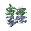


 PDBj
PDBj


 Assembly
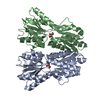

 Mass: 92.094 Da / Num. of mol.: 2 / Source method: obtained synthetically / Formula: C3H8O3
 Mass: 35.453 Da / Num. of mol.: 1 / Source method: obtained synthetically / Formula: Cl
 Mass: 18.015 Da / Num. of mol.: 253 / Source method: isolated from a natural source / Formula: H2O
 Sample preparation
 / Beamline: X29A / Wavelength: 0.979 Å
 Processing