 Movie
Movie Controller

[English] 日本語
 Yorodumi
- PDB-3ctd: Crystal structure of a putative AAA family ATPase from Prochloroc... -
+ Open data

- Basic information
| Entry | Database: PDB / ID: 3ctd | ||||||
|---|---|---|---|---|---|---|---|
| Title | Crystal structure of a putative AAA family ATPase from Prochlorococcus marinus subsp. pastoris | ||||||
 Components Components | Putative ATPase, AAA family | ||||||
 Keywords Keywords | STRUCTURAL GENOMICS / UNKNOWN FUNCTION / PSI-2 / Protein Structure Initiative / New York SGX Research Center for Structural Genomics / NYSGXRC / ATP-binding / Nucleotide-binding | ||||||
| Function / homology |  Function and homology information Function and homology informationfour-way junction helicase activity / DNA recombination / DNA replication / nucleotide binding / DNA repair / DNA bindingSimilarity search - Function | ||||||
| Biological species |  Prochlorococcus marinus subsp. pastoris str. CCMP1986 (bacteria) Prochlorococcus marinus subsp. pastoris str. CCMP1986 (bacteria) | ||||||
| Method | X-RAY DIFFRACTION / SYNCHROTRON / SAD / Resolution: 2.5 Å | ||||||
 Authors Authors | Bonanno, J.B. / Rutter, M. / Bain, K.T. / Lau, C. / Ozyurt, S. / Smith, D. / Wasserman, S. / Sauder, J.M. / Burley, S.K. / Almo, S.C. / New York SGX Research Center for Structural Genomics (NYSGXRC) | ||||||
 Citation Citation | Journal: To be Published Title: Crystal structure of a putative AAA family ATPase from Prochlorococcus marinus subsp. pastoris. Authors: Bonanno, J.B. / Rutter, M. / Bain, K.T. / Lau, C. / Ozyurt, S. / Smith, D. / Wasserman, S. / Sauder, J.M. / Burley, S.K. / Almo, S.C. | ||||||
| History |
|
- Structure visualization
| Structure viewer | Molecule: MolmilJmol/JSmol |
|---|
- Downloads & links
-Download
| PDBx/mmCIF format | 3ctd.cif.gz | 73.7 KB | Display | PDBx/mmCIF format |
|---|---|---|---|---|
| PDB format | pdb3ctd.ent.gz | 54.4 KB | Display | PDB format |
| PDBx/mmJSON format | 3ctd.json.gz | Tree view | PDBx/mmJSON format | |
| Others |  Other downloads Other downloads |
-Validation report
| Arichive directory | https://data.pdbj.org/pub/pdb/validation_reports/ct/3ctdftp://data.pdbj.org/pub/pdb/validation_reports/ct/3ctd | HTTPS FTP |
|---|
-Related structure data
| Similar structure data | |
|---|---|
| Other databases |


-Links
 PDBj
PDBj
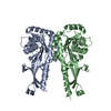
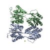
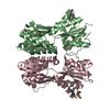
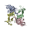
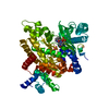
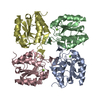
- Assembly
| Deposited unit | 
| ||||||||
|---|---|---|---|---|---|---|---|---|---|
| 1 | 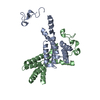
| ||||||||
| Unit cell |
|
-Components
| #1: Protein | Mass: 24161.311 Da / Num. of mol.: 2 / Fragment: Residues 228-429 / Mutation: L360M Source method: isolated from a genetically manipulated source Source: (gene. exp.) Prochlorococcus marinus subsp. pastoris str. CCMP1986 (bacteria)Species: Prochlorococcus marinus / Strain: CCMP 1378 / MED4 / Gene: yrvN, PMM0077 / Plasmid: Modified pET26 / Species (production host): Escherichia coli / Production host: Escherichia coli BL21(DE3) (bacteria) / Strain (production host): BL21(DE3) / References: UniProt: Q7V3J8#2: Water | ChemComp-HOH / | Water Mass: 18.015 Da / Num. of mol.: 58 / Source method: isolated from a natural source / Formula: H2O Mass: 18.015 Da / Num. of mol.: 58 / Source method: isolated from a natural source / Formula: H2O |
|---|
-Experimental details
-Experiment
| Experiment | Method: X-RAY DIFFRACTION / Number of used crystals: 1 |
|---|
- Sample preparation
| Crystal | Density Matthews: 2.08 Å3/Da / Density % sol: 40.85 % |
|---|---|
| Crystal grow | Temperature: 294 K / Method: vapor diffusion / pH: 7 Details: 100mM Hepes pH 7.0, 2% PEG 3350, 240mM Tri-sodium citrate dihydrate pH 7.0, VAPOR DIFFUSION, temperature 294K |
-Data collection
| Diffraction | Mean temperature: 100 K |
|---|---|
| Diffraction source | Source: SYNCHROTRON / Site: APS  / Beamline: 31-ID / Wavelength: 0.97958 Å / Beamline: 31-ID / Wavelength: 0.97958 Å |
| Detector | Type: MAR CCD 165 mm / Detector: CCD / Date: Mar 21, 2008 |
| Radiation | Monochromator: diamond / Protocol: SINGLE WAVELENGTH / Monochromatic (M) / Laue (L): M / Scattering type: x-ray |
| Radiation wavelength | Wavelength: 0.97958 Å / Relative weight: 1 |
| Reflection | Resolution: 2.5→39.559 Å / Num. all: 14631 / Num. obs: 14631 / % possible obs: 99.8 % / Observed criterion σ(F): 0 / Observed criterion σ(I): 0 / Redundancy: 8 % / Biso Wilson estimate: 37.6 Å2 / Rmerge(I) obs: 0.075 / Rsym value: 0.075 / Net I/σ(I): 22.7 |
| Reflection shell | Resolution: 2.5→2.64 Å / Redundancy: 8.3 % / Rmerge(I) obs: 0.123 / Mean I/σ(I) obs: 12.5 / Num. unique all: 2094 / Rsym value: 0.123 / % possible all: 100 |
- Processing
| Software |
| |||||||||||||||||||||||||||||||||||||||||||||||||||||||||||||||||
|---|---|---|---|---|---|---|---|---|---|---|---|---|---|---|---|---|---|---|---|---|---|---|---|---|---|---|---|---|---|---|---|---|---|---|---|---|---|---|---|---|---|---|---|---|---|---|---|---|---|---|---|---|---|---|---|---|---|---|---|---|---|---|---|---|---|---|
| Refinement | Method to determine structure: SAD / Resolution: 2.5→20 Å / Cor.coef. Fo:Fc: 0.924 / Cor.coef. Fo:Fc free: 0.875 / SU B: 8.107 / SU ML: 0.185 / Cross valid method: THROUGHOUT / ESU R: 0.414 / ESU R Free: 0.294 / Stereochemistry target values: MAXIMUM LIKELIHOOD
| |||||||||||||||||||||||||||||||||||||||||||||||||||||||||||||||||
| Solvent computation | Ion probe radii: 0.8 Å / Shrinkage radii: 0.8 Å / VDW probe radii: 1.4 Å / Solvent model: BABINET MODEL WITH MASK | |||||||||||||||||||||||||||||||||||||||||||||||||||||||||||||||||
| Displacement parameters | Biso mean: 40.801 Å2
| |||||||||||||||||||||||||||||||||||||||||||||||||||||||||||||||||
| Refinement step | Cycle: LAST / Resolution: 2.5→20 Å
| |||||||||||||||||||||||||||||||||||||||||||||||||||||||||||||||||
| Refine LS restraints |
| |||||||||||||||||||||||||||||||||||||||||||||||||||||||||||||||||
| LS refinement shell | Resolution: 2.5→2.564 Å / Total num. of bins used: 20
|
