Mass: 18.015 Da / Num. of mol.: 199 / Source method: isolated from a natural source / Formula: H2O
-
Details
Sequence details
THE CONSTRUCT WAS EXPRESSED WITH A PURIFICATION TAG MGSDKIHHHHHHENLYFQG. THE TAG WAS REMOVED WITH ...THE CONSTRUCT WAS EXPRESSED WITH A PURIFICATION TAG MGSDKIHHHHHHENLYFQG. THE TAG WAS REMOVED WITH TEV PROTEASE LEAVING ONLY A GLYCINE (0) FOLLOWED BY THE TARGET SEQUENCE.
-
Experimental details
-
Experiment
Experiment
Method: X-RAY DIFFRACTION / Number of used crystals: 1
-
Sample preparation
Crystal
Density Matthews: 2.48 Å3/Da / Density % sol: 50.48 %
Crystal grow
Temperature: 277 K / Method: vapor diffusion, sitting drop / pH: 6.5 Details: NANODROP, 2.0M (NH4)2SO4, 0.2M NaCl, 0.1M Cacodylate pH 6.5, VAPOR DIFFUSION, SITTING DROP, temperature 277K
Monochromator: Double crystal / Protocol: SINGLE WAVELENGTH / Monochromatic (M) / Laue (L): M / Scattering type: x-ray
Radiation wavelength
Wavelength: 0.97915 Å / Relative weight: 1
Reflection
Resolution: 2.15→29.735 Å / Num. obs: 20496 / % possible obs: 98.2 % / Observed criterion σ(I): -3 / Biso Wilson estimate: 29.356 Å2 / Rmerge(I) obs: 0.055 / Net I/σ(I): 10.33
Reflection shell
Resolution (Å)
Rmerge(I) obs
Mean I/σ(I) obs
Num. measured obs
Num. unique obs
Diffraction-ID
% possible all
2.15-2.23
0.322
2.4
7662
4013
1
96.5
2.23-2.32
0.242
3.2
7606
3952
1
97.6
2.32-2.42
0.194
3.9
7061
3663
1
97.9
2.42-2.55
0.168
4.6
7852
4075
1
98.7
2.55-2.71
0.122
6.1
7564
3907
1
98.9
2.71-2.92
0.093
7.8
7732
3989
1
98.6
2.92-3.21
0.061
11.2
7553
3883
1
99.1
3.21-3.67
0.037
16.9
7651
3931
1
98.7
3.67-4.61
0.026
22.8
7763
3968
1
98.8
4.61-29.735
0.026
24.1
7716
3938
1
97.4
-
Phasing
Phasing
Method: SAD
-
Processing
Software
Name
Version
Classification
NB
REFMAC
5.2.0019
refinement
PHENIX
refinement
SHELX
phasing
MolProbity
3beta29
modelbuilding
XSCALE
datascaling
PDB_EXTRACT
3.004
dataextraction
MAR345
CCD
datacollection
XDS
datareduction
SHELXD
phasing
autoSHARP
phasing
Refinement
Method to determine structure: SAD / Resolution: 2.15→29.735 Å / Cor.coef. Fo:Fc: 0.957 / Cor.coef. Fo:Fc free: 0.92 / SU B: 8.559 / SU ML: 0.118 / TLS residual ADP flag: LIKELY RESIDUAL / Cross valid method: THROUGHOUT / σ(F): 0 / ESU R: 0.216 / ESU R Free: 0.19 Stereochemistry target values: MAXIMUM LIKELIHOOD WITH PHASES Details: 1. HYDROGENS HAVE BEEN ADDED IN THE RIDING POSITIONS. 2. A MET-INHIBITION PROTOCOL WAS USED FOR SELENOMETHIONINE INCORPORATION DURING PROTEIN EXPRESSION. THE OCCUPANCY OF THE SE ATOMS IN THE ...Details: 1. HYDROGENS HAVE BEEN ADDED IN THE RIDING POSITIONS. 2. A MET-INHIBITION PROTOCOL WAS USED FOR SELENOMETHIONINE INCORPORATION DURING PROTEIN EXPRESSION. THE OCCUPANCY OF THE SE ATOMS IN THE MSE RESIDUES WAS REDUCED TO 0.75 FOR THE REDUCED SCATTERING POWER DUE TO PARTIAL S-MET INCORPORATION. 3. ATOM RECORD CONTAINS RESIDUAL B FACTORS ONLY. 4. ZN IS MODELED BASED ON AN X-RAY FLOURESCENCE SCAN, ANOMALOUS DIFFERENCE FOURIERS, AND COORDINATION GEOMETRY. 5. NA, CL, SO4, AND EDO ARE MODELED BASED ON CRYSTALLIZATION AND CRYO CONDITIONS.
Rfactor
Num. reflection
% reflection
Selection details
Rfree
0.228
1050
5.1 %
RANDOM
Rwork
0.168
-
-
-
obs
0.171
20494
99.64 %
-
Solvent computation
Ion probe radii: 0.8 Å / Shrinkage radii: 0.8 Å / VDW probe radii: 1.2 Å / Solvent model: MASK
Displacement parameters
Biso mean: 26.368 Å2
Baniso -1
Baniso -2
Baniso -3
1-
-0.56 Å2
0 Å2
-0.19 Å2
2-
-
1.11 Å2
0 Å2
3-
-
-
-0.63 Å2
Refinement step
Cycle: LAST / Resolution: 2.15→29.735 Å
Protein
Nucleic acid
Ligand
Solvent
Total
Num. atoms
2531
0
52
199
2782
Refine LS restraints
Refine-ID
Type
Dev ideal
Dev ideal target
Number
X-RAY DIFFRACTION
r_bond_refined_d
0.01
0.022
2654
X-RAY DIFFRACTION
r_bond_other_d
0.001
0.02
1822
X-RAY DIFFRACTION
r_angle_refined_deg
1.174
1.962
3590
X-RAY DIFFRACTION
r_angle_other_deg
0.906
3
4386
X-RAY DIFFRACTION
r_dihedral_angle_1_deg
5.588
5
328
X-RAY DIFFRACTION
r_dihedral_angle_2_deg
36.156
23.088
136
X-RAY DIFFRACTION
r_dihedral_angle_3_deg
13.222
15
415
X-RAY DIFFRACTION
r_dihedral_angle_4_deg
14.064
15
27
X-RAY DIFFRACTION
r_chiral_restr
0.07
0.2
391
X-RAY DIFFRACTION
r_gen_planes_refined
0.004
0.02
2985
X-RAY DIFFRACTION
r_gen_planes_other
0.001
0.02
567
X-RAY DIFFRACTION
r_nbd_refined
0.206
0.2
562
X-RAY DIFFRACTION
r_nbd_other
0.196
0.2
1918
X-RAY DIFFRACTION
r_nbtor_refined
0.176
0.2
1263
X-RAY DIFFRACTION
r_nbtor_other
0.084
0.2
1381
X-RAY DIFFRACTION
r_xyhbond_nbd_refined
0.157
0.2
161
X-RAY DIFFRACTION
r_metal_ion_refined
0.011
0.2
1
X-RAY DIFFRACTION
r_symmetry_vdw_refined
0.157
0.2
18
X-RAY DIFFRACTION
r_symmetry_vdw_other
0.258
0.2
41
X-RAY DIFFRACTION
r_symmetry_hbond_refined
0.152
0.2
16
X-RAY DIFFRACTION
r_mcbond_it
1.35
3
1622
X-RAY DIFFRACTION
r_mcbond_other
0.321
3
646
X-RAY DIFFRACTION
r_mcangle_it
2.425
5
2590
X-RAY DIFFRACTION
r_scbond_it
4.455
8
1056
X-RAY DIFFRACTION
r_scangle_it
6.405
11
996
LS refinement shell
Resolution: 2.15→2.206 Å / Total num. of bins used: 20
Rfactor
Num. reflection
% reflection
Rfree
0.253
66
-
Rwork
0.188
1414
-
all
-
1480
-
obs
-
-
98.47 %
Refinement TLS params.
Method: refined / Origin x: 14.22 Å / Origin y: 30.193 Å / Origin z: 18.446 Å
11
12
13
21
22
23
31
32
33
T
-0.0741 Å2
0.0115 Å2
-0.0243 Å2
-
-0.0707 Å2
-0.0069 Å2
-
-
-0.1464 Å2
L
0.5622 °2
0.1701 °2
-0.3121 °2
-
0.229 °2
-0.2843 °2
-
-
0.7762 °2
S
-0.015 Å °
-0.0172 Å °
0.0014 Å °
-0.0377 Å °
0.035 Å °
-0.005 Å °
0.0433 Å °
0.0351 Å °
-0.02 Å °
+
About Yorodumi
-
News
-
Feb 9, 2022. New format data for meta-information of EMDB entries
New format data for meta-information of EMDB entries
Version 3 of the EMDB header file is now the official format.
The previous official version 1.9 will be removed from the archive.
In the structure databanks used in Yorodumi, some data are registered as the other names, "COVID-19 virus" and "2019-nCoV". Here are the details of the virus and the list of structure data.
Jan 31, 2019. EMDB accession codes are about to change! (news from PDBe EMDB page)
EMDB accession codes are about to change! (news from PDBe EMDB page)
The allocation of 4 digits for EMDB accession codes will soon come to an end. Whilst these codes will remain in use, new EMDB accession codes will include an additional digit and will expand incrementally as the available range of codes is exhausted. The current 4-digit format prefixed with “EMD-” (i.e. EMD-XXXX) will advance to a 5-digit format (i.e. EMD-XXXXX), and so on. It is currently estimated that the 4-digit codes will be depleted around Spring 2019, at which point the 5-digit format will come into force.
The EM Navigator/Yorodumi systems omit the EMD- prefix.
Related info.:Q: What is EMD? / ID/Accession-code notation in Yorodumi/EM Navigator
Yorodumi is a browser for structure data from EMDB, PDB, SASBDB, etc.
This page is also the successor to EM Navigator detail page, and also detail information page/front-end page for Omokage search.
The word "yorodu" (or yorozu) is an old Japanese word meaning "ten thousand". "mi" (miru) is to see.
Related info.:EMDB / PDB / SASBDB / Comparison of 3 databanks / Yorodumi Search / Aug 31, 2016. New EM Navigator & Yorodumi / Yorodumi Papers / Jmol/JSmol / Function and homology information / Changes in new EM Navigator and Yorodumi
 Movie
Movie Controller

 Yorodumi
 Open data
 Basic information
 Components
 Keywords
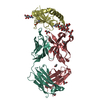
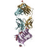
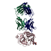
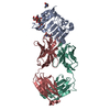
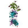
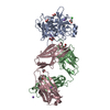
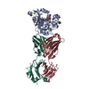
 TRANSFERASE / YP_614837.1 / Putative Aminoglycoside Phosphotransferase / Phosphotransferase enzyme family /
  Function and homology information
 Silicibacter sp. (bacteria)
 Authors
 Citation
 Structure visualization
 Downloads & links
 Other downloads
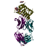


 PDBj
PDBj
 Assembly


 Mass: 65.409 Da / Num. of mol.: 1 / Source method: obtained synthetically / Formula: Zn
 Mass: 22.990 Da / Num. of mol.: 1 / Source method: obtained synthetically / Formula: Na
 Mass: 35.453 Da / Num. of mol.: 1 / Source method: obtained synthetically / Formula: Cl
 Mass: 96.063 Da / Num. of mol.: 1 / Source method: obtained synthetically / Formula: SO4
 Mass: 62.068 Da / Num. of mol.: 11 / Source method: obtained synthetically / Formula: C2H6O2
 Sample preparation
 / Beamline: BL9-2 / Wavelength: 0.97915 Å
 Processing