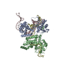
 Movie
Movie Controller


+ Open data

- Basic information
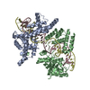
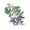
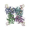
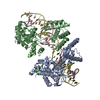
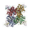
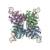
| Entry | Database: PDB / ID: 3crx | ||||||
|---|---|---|---|---|---|---|---|
| Title | CRE RECOMBINASE/DNA COMPLEX INTERMEDIATE I | ||||||
 Components Components |
| ||||||
 Keywords Keywords |  HYDROLASE / LIGASE/DNA / CRE RECOMBINASE / HOLLIDAY JUNCTION / RECOMBINATION / RECOMBINASE-DNA COMPLEX / LIGASE-DNA COMPLEX HYDROLASE / LIGASE/DNA / CRE RECOMBINASE / HOLLIDAY JUNCTION / RECOMBINATION / RECOMBINASE-DNA COMPLEX / LIGASE-DNA COMPLEX | ||||||
| Function / homology |  Function and homology information Function and homology information | ||||||
| Biological species |  Enterobacteria phage P1 (virus) Enterobacteria phage P1 (virus) | ||||||
| Method | X-RAY DIFFRACTION / SYNCHROTRON / DIFFERENCE FOURIER / Resolution: 2.5 Å | ||||||
 Authors Authors | Gopaul, D.N. / Guo, F. / Vanduyne, G.D. | ||||||
 Citation Citation | Journal: EMBO J. / Year: 1998 Title: Structure of the Holliday junction intermediate in Cre-loxP site-specific recombination. Authors: Gopaul, D.N. / Guo, F. / Van Duyne, G.D. #1: Journal: Nature / Year: 1997Title: Structure of Cre Recombinase Complexed with DNA in a Site-Specific Recombination Synapse Authors: Guo, F. / Gopaul, D.N. / Van Duyne, G.D. | ||||||
| History |
|
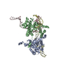
- Structure visualization
| Structure viewer | Molecule: MolmilJmol/JSmol |
|---|
- Downloads & links
-Download
| PDBx/mmCIF format | 3crx.cif.gz | 193.8 KB | Display | PDBx/mmCIF format |
|---|---|---|---|---|
| PDB format | pdb3crx.ent.gz | 152.9 KB | Display | PDB format |
| PDBx/mmJSON format | 3crx.json.gz | Tree view | PDBx/mmJSON format | |
| Others |  Other downloads Other downloads |
-Validation report
| Arichive directory | https://data.pdbj.org/pub/pdb/validation_reports/cr/3crxftp://data.pdbj.org/pub/pdb/validation_reports/cr/3crx | HTTPS FTP |
|---|
-Related structure data
| Related structure data | 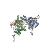 2crxC 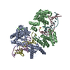 1crxS S: Starting model for refinement C: citing same article ( |
|---|---|
| Similar structure data |
-Links
 PDBj
PDBj


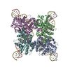
- Assembly
| Deposited unit | 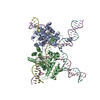
| ||||||||||
|---|---|---|---|---|---|---|---|---|---|---|---|
| 1 | 
| ||||||||||
| Unit cell |
|
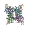
-Components
-DNA chain , 4 types, 4 molecules CDEF
| #1: DNA chain | Mass: 10758.980 Da / Num. of mol.: 1 / Source method: obtained synthetically |
|---|---|
| #2: DNA chain | Mass: 10718.956 Da / Num. of mol.: 1 / Source method: obtained synthetically |
| #3: DNA chain | Mass: 10758.980 Da / Num. of mol.: 1 / Source method: obtained synthetically |
| #4: DNA chain | Mass: 10799.004 Da / Num. of mol.: 1 / Source method: obtained synthetically |
-Protein / Non-polymers , 2 types, 238 molecules AB
| #5: Protein | / CRE-HJ1 / RGBPP1 RECOMBINASE Mass: 38567.152 Da / Num. of mol.: 2 / Mutation: R173K Source method: isolated from a genetically manipulated source Source: (gene. exp.) Enterobacteria phage P1 (virus) / Genus: P1-like viruses / Plasmid: PET21A / Species (production host): Escherichia coli / Production host:  Escherichia coli BL21(DE3) (bacteria) / Strain (production host): BL21 (DE3) / References: UniProt: P06956 Escherichia coli BL21(DE3) (bacteria) / Strain (production host): BL21 (DE3) / References: UniProt: P06956#6: Water | ChemComp-HOH / | WaterMass: 18.015 Da / Num. of mol.: 236 / Source method: isolated from a natural source / Formula: H2O |
|---|
-Details
| Compound details | THE STRUCTURE REPRESENTED IN THIS ENTRY CONTAINS TWO CRE MOLECULES AND FOUR DNA STRANDS. THE DNA IN ...THE STRUCTURE REPRESENTE |
|---|
-Experimental details
-Experiment
| Experiment | Method: X-RAY DIFFRACTION / Number of used crystals: 1 |
|---|
- Sample preparation
| Crystal | Density Matthews: 2.41 Å3/Da / Density % sol: 50 % | ||||||||||||||||||||||||||||||||||||||||||||||||||||||||||||||||||||||||||||||
|---|---|---|---|---|---|---|---|---|---|---|---|---|---|---|---|---|---|---|---|---|---|---|---|---|---|---|---|---|---|---|---|---|---|---|---|---|---|---|---|---|---|---|---|---|---|---|---|---|---|---|---|---|---|---|---|---|---|---|---|---|---|---|---|---|---|---|---|---|---|---|---|---|---|---|---|---|---|---|---|
| Crystal grow | pH: 5 / Details: 50MM ACETATE PH 5.0, 26% MPD, 80MM MGCL2 | ||||||||||||||||||||||||||||||||||||||||||||||||||||||||||||||||||||||||||||||
| Crystal grow | *PLUS Temperature: 4 ℃ / pH: 5.7 / Method: vapor diffusion, hanging drop | ||||||||||||||||||||||||||||||||||||||||||||||||||||||||||||||||||||||||||||||
| Components of the solutions | *PLUS
|
-Data collection
| Diffraction | Mean temperature: 100 K |
|---|---|
| Diffraction source | Source: SYNCHROTRON / Site: NSLS  / Beamline: X25 / Wavelength: 0.9 / Beamline: X25 / Wavelength: 0.9 |
| Detector | Type: MARRESEARCH / Detector: IMAGE PLATE / Date: May 15, 1997 / Details: MIRRORS |
| Radiation | Monochromator: SI / Protocol: SINGLE WAVELENGTH / Monochromatic (M) / Laue (L): M / Scattering type: x-ray |
| Radiation wavelength | Wavelength: 0.9 Å / Relative weight: 1 |
| Reflection | Resolution: 2.7→48 Å / Num. obs: 38807 / % possible obs: 99 % / Observed criterion σ(I): -3 / Redundancy: 4.7 % / Biso Wilson estimate: 65.5 Å2 / Rsym value: 0.072 / Net I/σ(I): 20 |
| Reflection shell | Resolution: 2.7→2.78 Å / Redundancy: 4.4 % / Mean I/σ(I) obs: 5 / Rsym value: 0.329 / % possible all: 93.2 |
| Reflection | *PLUS Rmerge(I) obs: 0.072 |
| Reflection shell | *PLUS % possible obs: 93.2 % / Rmerge(I) obs: 0.329 |
- Processing
| Software |
| ||||||||||||||||||||||||||||||||||||||||||||||||||||||||||||||||||||||||||||||||
|---|---|---|---|---|---|---|---|---|---|---|---|---|---|---|---|---|---|---|---|---|---|---|---|---|---|---|---|---|---|---|---|---|---|---|---|---|---|---|---|---|---|---|---|---|---|---|---|---|---|---|---|---|---|---|---|---|---|---|---|---|---|---|---|---|---|---|---|---|---|---|---|---|---|---|---|---|---|---|---|---|---|
| Refinement | Method to determine structure: DIFFERENCE FOURIER Starting model: 1CRX Resolution: 2.5→27.3 Å / Rfactor Rfree error: 0.017 / Data cutoff high absF: 1000000 / Data cutoff low absF: 0.001 / Isotropic thermal model: RESTRAINED / Cross valid method: THROUGHOUT / σ(F): 2
| ||||||||||||||||||||||||||||||||||||||||||||||||||||||||||||||||||||||||||||||||
| Displacement parameters | Biso mean: 58.2 Å2 | ||||||||||||||||||||||||||||||||||||||||||||||||||||||||||||||||||||||||||||||||
| Refinement step | Cycle: LAST / Resolution: 2.5→27.3 Å
| ||||||||||||||||||||||||||||||||||||||||||||||||||||||||||||||||||||||||||||||||
| Refine LS restraints |
| ||||||||||||||||||||||||||||||||||||||||||||||||||||||||||||||||||||||||||||||||
| LS refinement shell | Resolution: 2.5→2.61 Å / Rfactor Rfree error: 0.017 / Total num. of bins used: 8
| ||||||||||||||||||||||||||||||||||||||||||||||||||||||||||||||||||||||||||||||||
| Xplor file |
| ||||||||||||||||||||||||||||||||||||||||||||||||||||||||||||||||||||||||||||||||
| Software | *PLUS Name: X-PLOR / Version: 3.851 / Classification: refinement | ||||||||||||||||||||||||||||||||||||||||||||||||||||||||||||||||||||||||||||||||
| Refinement | *PLUS | ||||||||||||||||||||||||||||||||||||||||||||||||||||||||||||||||||||||||||||||||
| Solvent computation | *PLUS | ||||||||||||||||||||||||||||||||||||||||||||||||||||||||||||||||||||||||||||||||
| Displacement parameters | *PLUS | ||||||||||||||||||||||||||||||||||||||||||||||||||||||||||||||||||||||||||||||||
| Refine LS restraints | *PLUS
| ||||||||||||||||||||||||||||||||||||||||||||||||||||||||||||||||||||||||||||||||
| LS refinement shell | *PLUS Rfactor obs: 0.344 |
