 Movie
Movie Controller

[English] 日本語
 Yorodumi
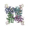
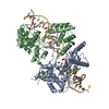
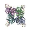
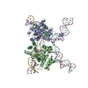
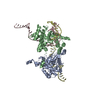
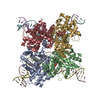
- PDB-1q3u: Crystal structure of a wild-type Cre recombinase-loxP synapse: pr... -
+ Open data

- Basic information
| Entry | Database: PDB / ID: 1q3u | ||||||
|---|---|---|---|---|---|---|---|
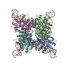
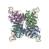
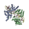
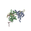
| Title | Crystal structure of a wild-type Cre recombinase-loxP synapse: pre-cleavage complex | ||||||
 Components Components |
| ||||||
 Keywords Keywords | REPLICATION/DNA / Cre /  recombinase / DNA-protein complex / crystal / REPLICATION-DNA COMPLEX recombinase / DNA-protein complex / crystal / REPLICATION-DNA COMPLEX | ||||||
| Function / homology |  Function and homology information Function and homology information | ||||||
| Biological species |  Enterobacteria phage P1 (virus) Enterobacteria phage P1 (virus) | ||||||
| Method | X-RAY DIFFRACTION / SYNCHROTRON / MOLECULAR REPLACEMENT / Resolution: 2.9 Å | ||||||
 Authors Authors | Ennifar, E. / Meyer, J.E.W. / Buchholz, F. / Stewart, A.F. / Suck, D. | ||||||
 Citation Citation | Journal: Nucleic Acids Res. / Year: 2003 Title: Crystal structure of a wild-type Cre recombinase-loxP synapse reveals a novel spacer conformation suggesting an alternative mechanism for DNA cleavage activation Authors: Ennifar, E. / Meyer, J.E.W. / Buchholz, F. / Stewart, A.F. / Suck, D. | ||||||
| History |
|
- Structure visualization
| Structure viewer | Molecule: MolmilJmol/JSmol |
|---|
- Downloads & links
-Download
| PDBx/mmCIF format | 1q3u.cif.gz | 342.4 KB | Display | PDBx/mmCIF format |
|---|---|---|---|---|
| PDB format | pdb1q3u.ent.gz | 270.9 KB | Display | PDB format |
| PDBx/mmJSON format | 1q3u.json.gz | Tree view | PDBx/mmJSON format | |
| Others |  Other downloads Other downloads |
-Validation report
| Arichive directory | https://data.pdbj.org/pub/pdb/validation_reports/q3/1q3uftp://data.pdbj.org/pub/pdb/validation_reports/q3/1q3u | HTTPS FTP |
|---|
-Related structure data
| Related structure data |  1nzbC  1ouqC  1q3vC 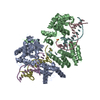 1crxS C: citing same article ( S: Starting model for refinement |
|---|---|
| Similar structure data |
-Links
 PDBj
PDBj


- Assembly
| Deposited unit | 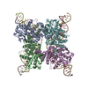
| ||||||||||
|---|---|---|---|---|---|---|---|---|---|---|---|
| 1 |
| ||||||||||
| Unit cell |
| ||||||||||
| Details | dimeric Cre recombinase bound to a double stranded DNA loxP site |
-Components
-DNA chain , 2 types, 4 molecules CGDH
| #1: DNA chain | Mass: 11364.330 Da / Num. of mol.: 2 / Source method: obtained synthetically / Details: First strand of loxP site #2: DNA chain | Mass: 11387.370 Da / Num. of mol.: 2 / Source method: obtained synthetically / Details: Second strand of loxP site |
|---|
-Protein , 1 types, 4 molecules ABEF
| #3: Protein | / GST-loxP-cre recombinase fusion protein / Retrofitting vector pRetroES Mass: 39066.715 Da / Num. of mol.: 4 Source method: isolated from a genetically manipulated source Source: (gene. exp.) Enterobacteria phage P1 (virus) / Genus: P1-like viruses / Plasmid: pGem / Production host:  Escherichia coli (E. coli) / References: UniProt: P06956 Escherichia coli (E. coli) / References: UniProt: P06956 |
|---|
-Non-polymers , 3 types, 15 molecules 


| #4: Chemical | Iodide Mass: 126.904 Da / Num. of mol.: 2 / Source method: obtained synthetically / Formula: I Mass: 126.904 Da / Num. of mol.: 2 / Source method: obtained synthetically / Formula: I#5: Chemical | ChemComp-MG /  Mass: 24.305 Da / Num. of mol.: 10 / Source method: obtained synthetically / Formula: Mg Mass: 24.305 Da / Num. of mol.: 10 / Source method: obtained synthetically / Formula: Mg#6: Water | ChemComp-HOH / | WaterMass: 18.015 Da / Num. of mol.: 3 / Source method: isolated from a natural source / Formula: H2O |
|---|
-Experimental details
-Experiment
| Experiment | Method: X-RAY DIFFRACTION / Number of used crystals: 1 |
|---|
- Sample preparation
| Crystal | Density Matthews: 4.58 Å3/Da / Density % sol: 72.9 % | ||||||||||||||||||||||||||||||||||||||||||||||||
|---|---|---|---|---|---|---|---|---|---|---|---|---|---|---|---|---|---|---|---|---|---|---|---|---|---|---|---|---|---|---|---|---|---|---|---|---|---|---|---|---|---|---|---|---|---|---|---|---|---|
| Crystal grow | Temperature: 293 K / Method: vapor diffusion, hanging drop / pH: 7.6 Details: NaCl, EDTA, Glycerol, HEPES, MgCl2, PEG 2000, MME, pH 7.6, VAPOR DIFFUSION, HANGING DROP, temperature 293K | ||||||||||||||||||||||||||||||||||||||||||||||||
| Components of the solutions |
| ||||||||||||||||||||||||||||||||||||||||||||||||
| Crystal grow | *PLUS Method: unknown | ||||||||||||||||||||||||||||||||||||||||||||||||
| Components of the solutions | *PLUS
|
-Data collection
| Diffraction | Mean temperature: 100 K |
|---|---|
| Diffraction source | Source: SYNCHROTRON / Site: ESRF  / Beamline: ID29 / Wavelength: 1 Å / Beamline: ID29 / Wavelength: 1 Å |
| Detector | Type: ADSC QUANTUM 4 / Detector: CCD |
| Radiation | Monochromator: crystal / Protocol: SINGLE WAVELENGTH / Monochromatic (M) / Laue (L): M / Scattering type: x-ray |
| Radiation wavelength | Wavelength: 1 Å / Relative weight: 1 |
| Reflection | Resolution: 2.8→30 Å / % possible obs: 97.4 % / Observed criterion σ(F): 0 / Observed criterion σ(I): 0 / Biso Wilson estimate: 57.8 Å2 / Rsym value: 0.073 |
| Reflection shell | Highest resolution: 2.8 Å / Rsym value: 0.318 |
| Reflection | *PLUS Highest resolution: 2.8 Å / Rmerge(I) obs: 0.073 |
| Reflection shell | *PLUS % possible obs: 94.6 % / Rmerge(I) obs: 0.318 |
- Processing
| Software |
| ||||||||||||||||||||||||||||||||||||
|---|---|---|---|---|---|---|---|---|---|---|---|---|---|---|---|---|---|---|---|---|---|---|---|---|---|---|---|---|---|---|---|---|---|---|---|---|---|
| Refinement | Method to determine structure: MOLECULAR REPLACEMENT Starting model: 1CRX Resolution: 2.9→20.03 Å / Rfactor Rfree error: 0.004 / Isotropic thermal model: RESTRAINED / Cross valid method: THROUGHOUT / σ(F): 2
| ||||||||||||||||||||||||||||||||||||
| Solvent computation | Solvent model: FLAT MODEL / Bsol: 37.4354 Å2 / ksol: 0.325868 e/Å3 | ||||||||||||||||||||||||||||||||||||
| Displacement parameters | Biso mean: 57.8 Å2
| ||||||||||||||||||||||||||||||||||||
| Refine analyze | Luzzati coordinate error free: 0.42 Å / Luzzati sigma a free: 0.41 Å | ||||||||||||||||||||||||||||||||||||
| Refinement step | Cycle: LAST / Resolution: 2.9→20.03 Å
| ||||||||||||||||||||||||||||||||||||
| Refine LS restraints |
| ||||||||||||||||||||||||||||||||||||
| LS refinement shell | Resolution: 2.9→3.08 Å / Rfactor Rfree error: 0.023 / Total num. of bins used: 6
| ||||||||||||||||||||||||||||||||||||
| Xplor file |
| ||||||||||||||||||||||||||||||||||||
| Refinement | *PLUS Highest resolution: 2.8 Å / Rfactor Rfree: 0.252 / Rfactor Rwork: 0.23 | ||||||||||||||||||||||||||||||||||||
| Solvent computation | *PLUS | ||||||||||||||||||||||||||||||||||||
| Displacement parameters | *PLUS | ||||||||||||||||||||||||||||||||||||
| Refine LS restraints | *PLUS
|
