 Movie
Movie Controller

[English] 日本語
 Yorodumi
- PDB-3cr7: Crystal structure of N-terminal truncation of APS Kinase from Pen... -
+ Open data

- Basic information
| Entry | Database: PDB / ID: 3cr7 | ||||||
|---|---|---|---|---|---|---|---|
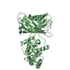
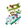
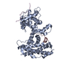
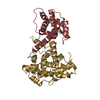
| Title | Crystal structure of N-terminal truncation of APS Kinase from Penicillium chrysogenum: Ternary structure with ADP and PAPS | ||||||
 Components Components | Adenylyl-sulfate kinase | ||||||
 Keywords Keywords | TRANSFERASE / APS kinase / Adenylylsulfate kinase / sulfate metabolism / Nucleotide 2 kinase / Amino-acid biosynthesis / ATP-binding / Cysteine biosynthesis / Methionine biosynthesis / Nucleotide-binding / Phosphoprotein | ||||||
| Function / homology |  Function and homology informationadenylyl-sulfate kinase / adenylylsulfate kinase activity / sulfate assimilation / hydrogen sulfide biosynthetic process / cysteine biosynthetic process / methionine biosynthetic process / phosphorylation / ATP binding / cytosol Function and homology informationadenylyl-sulfate kinase / adenylylsulfate kinase activity / sulfate assimilation / hydrogen sulfide biosynthetic process / cysteine biosynthetic process / methionine biosynthetic process / phosphorylation / ATP binding / cytosolSimilarity search - Function | ||||||
| Biological species |  Penicillium chrysogenum (fungus) Penicillium chrysogenum (fungus) | ||||||
| Method | X-RAY DIFFRACTION / SYNCHROTRON / MOLECULAR REPLACEMENT / molecular replacement / Resolution: 2.5 Å | ||||||
 Authors Authors | Gay, S.C. / Segel, I.H. / Fisher, A.J. | ||||||
 Citation Citation | Journal: To be Published Title: Truncated APS kinase from Pencillium chrysogenum: Insight into the function of the N-terminal helix Authors: Gay, S.C. / Segel, I.H. / Fisher, A.J. | ||||||
| History |
|
- Structure visualization
| Structure viewer | Molecule: MolmilJmol/JSmol |
|---|
- Downloads & links
-Download
| PDBx/mmCIF format | 3cr7.cif.gz | 168 KB | Display | PDBx/mmCIF format |
|---|---|---|---|---|
| PDB format | pdb3cr7.ent.gz | 132.3 KB | Display | PDB format |
| PDBx/mmJSON format | 3cr7.json.gz | Tree view | PDBx/mmJSON format | |
| Others |  Other downloads Other downloads |
-Validation report
| Arichive directory | https://data.pdbj.org/pub/pdb/validation_reports/cr/3cr7ftp://data.pdbj.org/pub/pdb/validation_reports/cr/3cr7 | HTTPS FTP |
|---|
-Related structure data
| Related structure data | 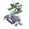 1m7gS S: Starting model for refinement |
|---|---|
| Similar structure data |


-Links
 PDBj
PDBj
- Assembly
| Deposited unit | 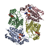
| ||||||||
|---|---|---|---|---|---|---|---|---|---|
| 1 | 
| ||||||||
| 2 | 
| ||||||||
| Unit cell |
|
-Components
| #1: Protein | / APS kinase / Adenosine-5'-phosphosulfate kinase / ATP adenosine-5'-phosphosulfate 3'-phosphotransferase Mass: 22354.123 Da / Num. of mol.: 4 / Fragment: UNP residues 23-211 Source method: isolated from a genetically manipulated source Source: (gene. exp.) Penicillium chrysogenum (fungus) / Strain: ATTC 24791 / Plasmid: pET22b / Production host:  Escherichia coli (E. coli) / Strain (production host): BL21(DE3) / References: UniProt: Q12657, adenylyl-sulfate kinase Escherichia coli (E. coli) / Strain (production host): BL21(DE3) / References: UniProt: Q12657, adenylyl-sulfate kinase#2: Chemical | Chloride  Mass: 35.453 Da / Num. of mol.: 3 / Source method: obtained synthetically / Formula: Cl Mass: 35.453 Da / Num. of mol.: 3 / Source method: obtained synthetically / Formula: Cl#3: Chemical | ChemComp-ADP / Adenosine diphosphate  Mass: 427.201 Da / Num. of mol.: 4 / Source method: obtained synthetically / Formula: C10H15N5O10P2 / Comment: ADP, energy-carrying molecule*YM Mass: 427.201 Da / Num. of mol.: 4 / Source method: obtained synthetically / Formula: C10H15N5O10P2 / Comment: ADP, energy-carrying molecule*YM#4: Chemical | 3'-Phosphoadenosine-5'-phosphosulfate  Mass: 507.264 Da / Num. of mol.: 2 / Source method: obtained synthetically / Formula: C10H15N5O13P2S Mass: 507.264 Da / Num. of mol.: 2 / Source method: obtained synthetically / Formula: C10H15N5O13P2S#5: Water | ChemComp-HOH / | Water Mass: 18.015 Da / Num. of mol.: 495 / Source method: isolated from a natural source / Formula: H2O Mass: 18.015 Da / Num. of mol.: 495 / Source method: isolated from a natural source / Formula: H2O |
|---|
-Experimental details
-Experiment
| Experiment | Method: X-RAY DIFFRACTION / Number of used crystals: 1 |
|---|
- Sample preparation
| Crystal | Density Matthews: 2.53 Å3/Da / Density % sol: 51.37 % |
|---|---|
| Crystal grow | Temperature: 293 K / Method: vapor diffusion, hanging drop / pH: 6.5 Details: bis tris, PEG 3350, ammonium acetate, pH 6.5, VAPOR DIFFUSION, HANGING DROP, temperature 293K |
-Data collection
| Diffraction | Mean temperature: 100 K |
|---|---|
| Diffraction source | Source: SYNCHROTRON / Site: SSRL  / Beamline: BL1-5 / Wavelength: 0.9194 Å / Beamline: BL1-5 / Wavelength: 0.9194 Å |
| Detector | Type: ADSC QUANTUM 315 / Detector: CCD / Date: Mar 29, 2007 / Details: Double-crystal monochromator |
| Radiation | Monochromator: Double Si(111) crystal, paralell / Protocol: SINGLE WAVELENGTH / Monochromatic (M) / Laue (L): M / Scattering type: x-ray |
| Radiation wavelength | Wavelength: 0.9194 Å / Relative weight: 1 |
| Reflection | Resolution: 2.5→31.718 Å / Num. all: 31582 / Num. obs: 31427 / % possible obs: 99.6 % / Observed criterion σ(F): -3 / Observed criterion σ(I): -3 / Redundancy: 3.6 % / Rmerge(I) obs: 0.084 / Rsym value: 0.084 / Net I/σ(I): 7.5 |
| Reflection shell | Resolution: 2.5→2.56 Å / Redundancy: 3.6 % / Rmerge(I) obs: 0.486 / Mean I/σ(I) obs: 1.5 / Num. measured all: 8337 / Num. unique all: 2290 / Rsym value: 0.486 / % possible all: 100 |
-Phasing
| Phasing | Method: molecular replacement | ||||||
|---|---|---|---|---|---|---|---|
| Phasing MR |
|
- Processing
| Software |
| |||||||||||||||||||||||||||||||||||||||||||||||||||||||||||||||||||||||||||||||||||||||||||||||||||||||||||||||||||||||
|---|---|---|---|---|---|---|---|---|---|---|---|---|---|---|---|---|---|---|---|---|---|---|---|---|---|---|---|---|---|---|---|---|---|---|---|---|---|---|---|---|---|---|---|---|---|---|---|---|---|---|---|---|---|---|---|---|---|---|---|---|---|---|---|---|---|---|---|---|---|---|---|---|---|---|---|---|---|---|---|---|---|---|---|---|---|---|---|---|---|---|---|---|---|---|---|---|---|---|---|---|---|---|---|---|---|---|---|---|---|---|---|---|---|---|---|---|---|---|---|---|
| Refinement | Method to determine structure: MOLECULAR REPLACEMENT Starting model: poly alanine model of pdb entry 1M7G A chain Resolution: 2.5→31.715 Å / Isotropic thermal model: isotropic / σ(F): 0.01 / Stereochemistry target values: ML
| |||||||||||||||||||||||||||||||||||||||||||||||||||||||||||||||||||||||||||||||||||||||||||||||||||||||||||||||||||||||
| Solvent computation | Shrinkage radii: 0.9 Å / VDW probe radii: 1.11 Å / Solvent model: FLAT BULK SOLVENT MODEL | |||||||||||||||||||||||||||||||||||||||||||||||||||||||||||||||||||||||||||||||||||||||||||||||||||||||||||||||||||||||
| Displacement parameters | Biso max: 0 Å2 / Biso mean: 37.49 Å2 / Biso min: 391.05 Å2
| |||||||||||||||||||||||||||||||||||||||||||||||||||||||||||||||||||||||||||||||||||||||||||||||||||||||||||||||||||||||
| Refinement step | Cycle: LAST / Resolution: 2.5→31.715 Å
| |||||||||||||||||||||||||||||||||||||||||||||||||||||||||||||||||||||||||||||||||||||||||||||||||||||||||||||||||||||||
| Refine LS restraints |
| |||||||||||||||||||||||||||||||||||||||||||||||||||||||||||||||||||||||||||||||||||||||||||||||||||||||||||||||||||||||
| LS refinement shell | Refine-ID: X-RAY DIFFRACTION / Total num. of bins used: 16
|
