 Movie
Movie Controller


+ Open data

- Basic information
| Entry | Database: PDB / ID: 2yvu | ||||||
|---|---|---|---|---|---|---|---|
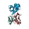
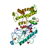
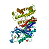
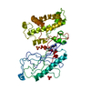
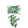
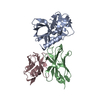
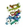
| Title | Crystal structure of APE1195 | ||||||
 Components Components | Probable adenylyl-sulfate kinase | ||||||
 Keywords Keywords |  TRANSFERASE / KINASE / Structural Genomics / NPPSFA / National Project on Protein Structural and Functional Analyses / RIKEN Structural Genomics/Proteomics Initiative / RSGI TRANSFERASE / KINASE / Structural Genomics / NPPSFA / National Project on Protein Structural and Functional Analyses / RIKEN Structural Genomics/Proteomics Initiative / RSGI | ||||||
| Function / homology |  Function and homology informationadenylyl-sulfate kinase / adenylylsulfate kinase activity / sulfate assimilation / hydrogen sulfide biosynthetic process / ATP binding Function and homology informationadenylyl-sulfate kinase / adenylylsulfate kinase activity / sulfate assimilation / hydrogen sulfide biosynthetic process / ATP bindingSimilarity search - Function | ||||||
| Biological species |   Aeropyrum pernix (archaea) Aeropyrum pernix (archaea) | ||||||
| Method | X-RAY DIFFRACTION / SYNCHROTRON / MOLECULAR REPLACEMENT / Resolution: 2.1 Å | ||||||
 Authors Authors | Ishii, R. / Bessho, Y. / Yokoyama, S. / RIKEN Structural Genomics/Proteomics Initiative (RSGI) | ||||||
 Citation Citation | Journal: To be published Title: Crystal structure of APE1195 Authors: Ishii, R. / Bessho, Y. / Yokoyama, S. | ||||||
| History |
| ||||||
| Remark 999 | SEQUENCE UNIPROT Q9YCR6 was updated on 2007-06-26. The author constructed the plasmid following the ...SEQUENCE UNIPROT Q9YCR6 was updated on 2007-06-26. The author constructed the plasmid following the old database Q9YCR6 including the first 4 residues MET GLN ALA LEU. |
- Structure visualization
| Structure viewer | Molecule: MolmilJmol/JSmol |
|---|
- Downloads & links
-Download
| PDBx/mmCIF format | 2yvu.cif.gz | 82.8 KB | Display | PDBx/mmCIF format |
|---|---|---|---|---|
| PDB format | pdb2yvu.ent.gz | 63 KB | Display | PDB format |
| PDBx/mmJSON format | 2yvu.json.gz | Tree view | PDBx/mmJSON format | |
| Others |  Other downloads Other downloads |
-Validation report
| Arichive directory | https://data.pdbj.org/pub/pdb/validation_reports/yv/2yvuftp://data.pdbj.org/pub/pdb/validation_reports/yv/2yvu | HTTPS FTP |
|---|
-Related structure data
| Related structure data |  2gksS S: Starting model for refinement |
|---|---|
| Similar structure data | |
| Other databases |


-Links
 PDBj
PDBj
- Assembly
| Deposited unit | 
| ||||||||
|---|---|---|---|---|---|---|---|---|---|
| 1 |
| ||||||||
| Unit cell |
|
-Components
| #1: Protein | Mass: 21123.367 Da / Num. of mol.: 2 Source method: isolated from a genetically manipulated source Source: (gene. exp.) Aeropyrum pernix (archaea) / Strain: K1 / Gene: cysC / Plasmid: pET-21a / Production host:  Escherichia coli (E. coli) / Strain (production host): BL21-CodonPlus(DE3)-RIL / References: UniProt: Q9YCR6, adenylyl-sulfate kinase Escherichia coli (E. coli) / Strain (production host): BL21-CodonPlus(DE3)-RIL / References: UniProt: Q9YCR6, adenylyl-sulfate kinase#2: Water | ChemComp-HOH / | Water Mass: 18.015 Da / Num. of mol.: 131 / Source method: isolated from a natural source / Formula: H2O Mass: 18.015 Da / Num. of mol.: 131 / Source method: isolated from a natural source / Formula: H2O |
|---|
-Experimental details
-Experiment
| Experiment | Method: X-RAY DIFFRACTION / Number of used crystals: 1 |
|---|
- Sample preparation
| Crystal | Density Matthews: 2.42 Å3/Da / Density % sol: 49.25 % |
|---|---|
| Crystal grow | Temperature: 293 K / Method: oil batch / pH: 7.6 Details: 15% (v/v) MPD, 0.1M HEPES-NaOH, 0.2M tri-Na Citr, pH 7.6, oil batch, temperature 293K |
-Data collection
| Diffraction | Mean temperature: 100 K |
|---|---|
| Diffraction source | Source: SYNCHROTRON / Site: SPring-8  / Beamline: BL26B2 / Wavelength: 1 Å / Beamline: BL26B2 / Wavelength: 1 Å |
| Detector | Type: RIGAKU JUPITER 210 / Detector: CCD / Date: Mar 28, 2007 |
| Radiation | Protocol: SINGLE WAVELENGTH / Monochromatic (M) / Laue (L): M / Scattering type: x-ray |
| Radiation wavelength | Wavelength: 1 Å / Relative weight: 1 |
| Reflection | Resolution: 2.1→50 Å / Num. all: 23374 / Num. obs: 23213 / % possible obs: 98.8 % / Observed criterion σ(I): -3 / Redundancy: 5.3 % / Biso Wilson estimate: 22.2 Å2 / Rmerge(I) obs: 0.045 / Net I/σ(I): 31.2 |
| Reflection shell | Resolution: 2.1→2.18 Å / Redundancy: 4.4 % / Rmerge(I) obs: 0.159 / Mean I/σ(I) obs: 5.4 / Num. unique all: 2117 / % possible all: 91.3 |
- Processing
| Software |
| ||||||||||||||||||||||||||||||||||||
|---|---|---|---|---|---|---|---|---|---|---|---|---|---|---|---|---|---|---|---|---|---|---|---|---|---|---|---|---|---|---|---|---|---|---|---|---|---|
| Refinement | Method to determine structure: MOLECULAR REPLACEMENT Starting model: PDB ENTRY 2GKS Resolution: 2.1→45.27 Å / Rfactor Rfree error: 0.007 / Data cutoff high absF: 1283584.86 / Data cutoff low absF: 0 / Isotropic thermal model: RESTRAINED / Cross valid method: THROUGHOUT / σ(F): 0 / Stereochemistry target values: Engh & Huber / Details: BULK SOLVENT MODEL USED
| ||||||||||||||||||||||||||||||||||||
| Solvent computation | Solvent model: FLAT MODEL / Bsol: 50.8713 Å2 / ksol: 0.35 e/Å3 | ||||||||||||||||||||||||||||||||||||
| Displacement parameters | Biso mean: 38.3 Å2
| ||||||||||||||||||||||||||||||||||||
| Refine analyze |
| ||||||||||||||||||||||||||||||||||||
| Refinement step | Cycle: LAST / Resolution: 2.1→45.27 Å
| ||||||||||||||||||||||||||||||||||||
| Refine LS restraints |
| ||||||||||||||||||||||||||||||||||||
| LS refinement shell | Resolution: 2.1→2.23 Å / Rfactor Rfree error: 0.02 / Total num. of bins used: 6
| ||||||||||||||||||||||||||||||||||||
| Xplor file |
|
