EXPERIMENTAL DETAILS EXPERIMENT TYPE : X-RAY SOLUTION SCATTERING DATA ACQUISITION ... EXPERIMENTAL DETAILS EXPERIMENT TYPE : X-RAY SOLUTION SCATTERING DATA ACQUISITION RADIATION/NEUTRON SOURCE :ESRF GRENOBLE SYNCHROTRON (Y/N) :Y BEAMLINE :ID2 BEAMLINE INSTRUMENT :NULL DETECTOR TYPE :FRELON CCD CAMERA DETECTOR MANUFACTURER DETAILS :NULL TEMPERATURE (KELVIN) :288 PH :7.5 NUMBER OF TIME FRAMES USED :10 PROTEIN CONCENTRATION RANGE (MG/ML) :0.32-0.90 SAMPLE BUFFER :140 MM NACL :12.5 MM NAHPO4 :0.5 MM EDTA :0.02% NA AZIDE DATA REDUCTION SOFTWARE :MULTICCD DATA ANALYSIS SOFTWARE :SCTPL7, GNOM GUINIER MEAN RADIUS OF GYRATION (NM) :8.13 SIGMA MEAN RADIUS OF GYRATION :0.10 R(XS-1) MEAN CROSS SECTIONAL RADII (NM) :4.22 R(XS-1) SIGMA MEAN CROSS SECTIONAL RADII :0.09 R(XS-2) MEAN CROSS SECTIONAL RADII (NM) :1.93 R(XS-2) SIGMA MEAN CROSS SECTIONAL RADII :0.03 P(R) PROTEIN LENGTH (NM) :26 P(R) SIGMA PROTEIN LENGTH :1 EXPERIMENTAL DETAILS EXPERIMENT TYPE : NEUTRON SOLUTION SCATTERING DATA ACQUISITION RADIATION/NEUTRON SOURCE :ISIS RUTHERFORD- :APPLETON LAB SYNCHROTRON (Y/N) :Y BEAMLINE :LOQ BEAMLINE INSTRUMENT :NULL DETECTOR TYPE :HE-3 ORDELA :DETECTOR DETECTOR MANUFACTURER DETAILS :NULL TEMPERATURE (KELVIN) :288 PH :7.5 NUMBER OF TIME FRAMES USED :1 PROTEIN CONCENTRATION RANGE (MG/ML) :1.94 SAMPLE BUFFER :140 MM NACL :12.5 MM NAHPO4 :0.5 MM EDTA :0.02% NA AZIDE DATA REDUCTION SOFTWARE :COLETTE DATA ANALYSIS SOFTWARE :SCTPL7, GNOM GUINIER MEAN RADIUS OF GYRATION (NM) :7.57 SIGMA MEAN RADIUS OF GYRATION :NULL R(XS-1) MEAN CROSS SECTIONAL RADII (NM) :NULL R(XS-1) SIGMA MEAN CROSS SECTIONAL RADII :NULL R(XS-2) MEAN CROSS SECTIONAL RADII (NM) :NULL R(XS-2) SIGMA MEAN CROSS SECTIONAL RADII :NULL P(R) PROTEIN LENGTH (NM) :24 P(R) SIGMA PROTEIN LENGTH :1
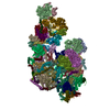
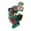
Secretory IgA2 is composed of four heavy chains (A, B, C, D) and four light chains (L, M, N, O), J chain (J) and secretory component (S)
-
Components
#1: Antibody
Immunoglobulinheavychain / / Coordinate model: Cα atoms only
Mass: 49922.031 Da / Num. of mol.: 4 / Source method: isolated from a natural source / Details: purified from human colostrum / Source: (natural) Homo sapiens (human)
#2: Protein
Secretorycomponent / / Coordinate model: Cα atoms only
Mass: 11850.517 Da / Num. of mol.: 1 / Fragment: Ig-like V-type domain 4 / Source method: isolated from a natural source / Details: purified from human colostrum / Source: (natural) Homo sapiens (human) / References: UniProt: P01833
#3: Antibody
Immunoglobulinlightchain / / Coordinate model: Cα atoms only
Mass: 23216.770 Da / Num. of mol.: 4 / Source method: isolated from a natural source / Details: purified from human colostrum / Source: (natural) Homo sapiens (human)
#4: Protein
Secretorycomponent / / Coordinate model: Cα atoms only
Mass: 64355.234 Da / Num. of mol.: 1 / Source method: isolated from a natural source / Details: purified from human colostrum / Source: (natural) Homo sapiens (human) / References: UniProt: P01833
Sequence details
THE PART OF A,B,C,D SEQUENCE BELONGS TO IGA VARIABLE HEAVY CHAIN.
-
Experimental details
-
Experiment
Experiment
Method: SOLUTION SCATTERING
-
Data collection
Diffraction
ID
Mean temperature (K)
Crystal-ID
1
288
1
2
288
1
Diffraction source
Source
Site
Beamline
ID
Wavelength (Å)
SYNCHROTRON
ESRF
ID2
1
1
SPALLATION SOURCE
ISIS
LOQ
2
2.0-10.0
Detector
Type
ID
Detector
Date
FRELON
1
CCD
Nov 1, 2001
3-He ORDELA
2
AREA DETECTOR
Jul 1, 2002
Radiation
ID
Monochromator
Protocol
Monochromatic (M) / Laue (L)
Scattering type
Wavelength-ID
2
TIMEOFFLIGHT
LAUE
L
neutron
1
1
MIRROR
SINGLEWAVELENGTH
M
x-ray
1
Radiation wavelength
ID
Wavelength (Å)
Relative weight
1
1
1
2
2
1
3
10
1
Soln scatter
Buffer name: 140 MM NACL 12.5 MM NAHPO4 0.5 MM EDTA 0.02% NA AZIDE / Data analysis software list: SCTPL7, GNOM / Protein length: 1 / Sample pH: 7.5 / Source class: Y / Temperature: 288 K
Type
ID
Conc. range (mg/ml)
Data reduction software list
Detector type
Max mean cross sectional radii gyration (nm)
Max mean cross sectional radii gyration esd (nm)
Mean guiner radius (nm)
Mean guiner radius esd (nm)
Min mean cross sectional radii gyration (nm)
Min mean cross sectional radii gyration esd (nm)
Num. of time frames
Source beamline
Source type
x-ray
1
0.32-0.90
MULTICCD
FRELONCCDCAMERA
1.93
0.03
8.13
0.1
4.22
0.09
10
ID2
ESRFGRENOBLE
neutron
2
1.94
COLETTE
HE-3 ORDELA DETECTOR
7.57
1
LOQ
ISIS RUTHERFORD- APPLETON LAB
-
Processing
Software
Name
Version
Classification
SCTPL7
modelbuilding
GNOM
modelbuilding
Insight II
II98
modelbuilding
COLETTE
(ISIS)
datascaling
SCTPL7
phasing
GNOM
phasing
Refinement
Method to determine structure: CONSTRAINED SCATTERING MODELLING Details: THE COORDINATES CONTAIN ONLY CA ATOMS.
Refinement step
Cycle: LAST
Protein
Nucleic acid
Ligand
Solvent
Total
Num. atoms
3395
0
0
0
3395
Soln scatter model
Num. of conformers submitted: 10 / Representative conformer: 1 / Software list: INSIGHT II, SCTPL7, GNOM
+
About Yorodumi
-
News
-
Feb 9, 2022. New format data for meta-information of EMDB entries
New format data for meta-information of EMDB entries
Version 3 of the EMDB header file is now the official format.
The previous official version 1.9 will be removed from the archive.
In the structure databanks used in Yorodumi, some data are registered as the other names, "COVID-19 virus" and "2019-nCoV". Here are the details of the virus and the list of structure data.
Jan 31, 2019. EMDB accession codes are about to change! (news from PDBe EMDB page)
EMDB accession codes are about to change! (news from PDBe EMDB page)
The allocation of 4 digits for EMDB accession codes will soon come to an end. Whilst these codes will remain in use, new EMDB accession codes will include an additional digit and will expand incrementally as the available range of codes is exhausted. The current 4-digit format prefixed with “EMD-” (i.e. EMD-XXXX) will advance to a 5-digit format (i.e. EMD-XXXXX), and so on. It is currently estimated that the 4-digit codes will be depleted around Spring 2019, at which point the 5-digit format will come into force.
The EM Navigator/Yorodumi systems omit the EMD- prefix.
Related info.:Q: What is EMD? / ID/Accession-code notation in Yorodumi/EM Navigator
Yorodumi is a browser for structure data from EMDB, PDB, SASBDB, etc.
This page is also the successor to EM Navigator detail page, and also detail information page/front-end page for Omokage search.
The word "yorodu" (or yorozu) is an old Japanese word meaning "ten thousand". "mi" (miru) is to see.
Related info.:EMDB / PDB / SASBDB / Comparison of 3 databanks / Yorodumi Search / Aug 31, 2016. New EM Navigator & Yorodumi / Yorodumi Papers / Jmol/JSmol / Function and homology information / Changes in new EM Navigator and Yorodumi
 Movie
Movie Controller


 Open data
 Basic information
 Components
 Keywords
 IMMUNE SYSTEM / Secretory IgA2 / Secretory IgA1 / IgA /
  Function and homology information
 Authors
 Citation
 Structure visualization
 Downloads & links
 Other downloads
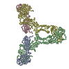


 PDBj
PDBj

 Assembly
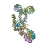

 Processing