 Movie
Movie Controller

[English] 日本語
 Yorodumi
- PDB-3clh: Crystal structure of 3-dehydroquinate synthase (DHQS)from Helicob... -
+ Open data

- Basic information
| Entry | Database: PDB / ID: 3clh | ||||||
|---|---|---|---|---|---|---|---|
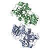
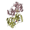
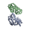
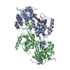
| Title | Crystal structure of 3-dehydroquinate synthase (DHQS)from Helicobacter pylori | ||||||
 Components Components | 3-dehydroquinate synthase | ||||||
 Keywords Keywords | LYASE / SHIKIMATE PATHWAY / AROMATIC AMINO ACID BIOSYNTHESIS / DHQS / Amino-acid biosynthesis / Cytoplasm / NAD | ||||||
| Function / homology |  Function and homology information3-dehydroquinate synthase / 3-dehydroquinate synthase activity / chorismate biosynthetic process / aromatic amino acid family biosynthetic process / nucleotide binding / metal ion binding / cytoplasm Function and homology information3-dehydroquinate synthase / 3-dehydroquinate synthase activity / chorismate biosynthetic process / aromatic amino acid family biosynthetic process / nucleotide binding / metal ion binding / cytoplasmSimilarity search - Function | ||||||
| Biological species |   Helicobacter pylori (bacteria) Helicobacter pylori (bacteria) | ||||||
| Method | X-RAY DIFFRACTION / MOLECULAR REPLACEMENT / Resolution: 2.4 Å | ||||||
 Authors Authors | Wang, W.C. / Liu, J.S. / Cheng, W.C. / Wang, H.J. / Chen, Y.C. | ||||||
 Citation Citation | Journal: Biochem.Biophys.Res.Commun. / Year: 2008 Title: Structure-based inhibitor discovery of Helicobacter pylori dehydroquinate synthase. Authors: Liu, J.S. / Cheng, W.C. / Wang, H.J. / Chen, Y.C. / Wang, W.C. | ||||||
| History |
|
- Structure visualization
| Structure viewer | Molecule: MolmilJmol/JSmol |
|---|
- Downloads & links
-Download
| PDBx/mmCIF format | 3clh.cif.gz | 139.3 KB | Display | PDBx/mmCIF format |
|---|---|---|---|---|
| PDB format | pdb3clh.ent.gz | 109 KB | Display | PDB format |
| PDBx/mmJSON format | 3clh.json.gz | Tree view | PDBx/mmJSON format | |
| Others |  Other downloads Other downloads |
-Validation report
| Arichive directory | https://data.pdbj.org/pub/pdb/validation_reports/cl/3clhftp://data.pdbj.org/pub/pdb/validation_reports/cl/3clh | HTTPS FTP |
|---|
-Related structure data
| Related structure data |  1nrxS S: Starting model for refinement |
|---|---|
| Similar structure data |


-Links
 PDBj
PDBj- Assembly
| Deposited unit | 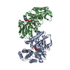
| ||||||||
|---|---|---|---|---|---|---|---|---|---|
| 1 |
| ||||||||
| Unit cell |
|
-Components
| #1: Protein | Mass: 39175.816 Da / Num. of mol.: 2 Source method: isolated from a genetically manipulated source Source: (gene. exp.) Helicobacter pylori (bacteria) / Gene: aroB / Plasmid: pQE30 / Production host: Escherichia coli (E. coli) / Strain (production host): JM109 / References: UniProt: P56081, 3-dehydroquinate synthase#2: Chemical |   Mass: 65.409 Da / Num. of mol.: 2 / Source method: obtained synthetically / Formula: Zn Mass: 65.409 Da / Num. of mol.: 2 / Source method: obtained synthetically / Formula: Zn#3: Chemical | Nicotinamide adenine dinucleotide  Mass: 663.425 Da / Num. of mol.: 2 / Source method: obtained synthetically / Formula: C21H27N7O14P2 / Comment: NAD*YM Mass: 663.425 Da / Num. of mol.: 2 / Source method: obtained synthetically / Formula: C21H27N7O14P2 / Comment: NAD*YM#4: Water | ChemComp-HOH / | Water Mass: 18.015 Da / Num. of mol.: 206 / Source method: isolated from a natural source / Formula: H2O Mass: 18.015 Da / Num. of mol.: 206 / Source method: isolated from a natural source / Formula: H2O |
|---|
-Experimental details
-Experiment
| Experiment | Method: X-RAY DIFFRACTION / Number of used crystals: 1 |
|---|
- Sample preparation
| Crystal | Density Matthews: 3 Å3/Da / Density % sol: 58.96 % |
|---|---|
| Crystal grow | Temperature: 298 K / Method: vapor diffusion, hanging drop / pH: 7.5 Details: 20mM NAD, 3.5M NaFormate, 0.1M Tris-HCl (pH 7.5), VAPOR DIFFUSION, HANGING DROP, temperature 298K |
-Data collection
| Diffraction source | Source: ROTATING ANODE / Type: RIGAKU RUH3R / Wavelength: 1.5418 Å |
|---|---|
| Detector | Type: RIGAKU RAXIS IV++ / Detector: IMAGE PLATE / Date: Feb 9, 2006 |
| Radiation | Protocol: SINGLE WAVELENGTH / Monochromatic (M) / Laue (L): M / Scattering type: x-ray |
| Radiation wavelength | Wavelength: 1.5418 Å / Relative weight: 1 |
| Reflection | Resolution: 2.4→79.31 Å / Num. all: 35600 / Num. obs: 35489 / % possible obs: 99.7 % |
| Reflection shell | Resolution: 2.4→2.49 Å |
- Processing
| Software |
| ||||||||||||||||||||||||||||||||||||
|---|---|---|---|---|---|---|---|---|---|---|---|---|---|---|---|---|---|---|---|---|---|---|---|---|---|---|---|---|---|---|---|---|---|---|---|---|---|
| Refinement | Method to determine structure: MOLECULAR REPLACEMENT Starting model: PDB ENTRY 1NRX Resolution: 2.4→30 Å / Cross valid method: THROUGHOUT / Stereochemistry target values: Engh & Huber
| ||||||||||||||||||||||||||||||||||||
| Refinement step | Cycle: LAST / Resolution: 2.4→30 Å
| ||||||||||||||||||||||||||||||||||||
| Refine LS restraints |
|
