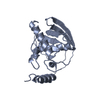
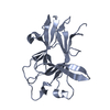
Entry Database : PDB / ID : 3ckhTitle Crystal structure of Eph A4 receptor Ephrin type-A receptor 4 Keywords / / / / / / / / / Function / homology Function Domain/homology Component
/ / / / / / / / / / / / / / / / / / / / / / / / / / / / / / / / / / / / / / / / / / / / / / / / / / / / / / / / / / / / / / / / / / / / / / / / / / / / / / / / / / / / / / / / / / / / / / / / / / / / / / / / / / / / / / / / / / / / / / / / / / / / / / / / / / / / / / / / / / Biological species Homo sapiens (human)Method / / / Resolution : 2.8 Å Authors Shi, J.H. / Song, J.X. Journal : J.Biol.Chem. / Year : 2008Title : Crystal Structure and NMR Binding Reveal That Two Small Molecule Antagonists Target the High Affinity Ephrin-binding Channel of the EphA4 Receptor.Authors : Qin, H. / Shi, J. / Noberini, R. / Pasquale, E.B. / Song, J. History Deposition Mar 15, 2008 Deposition site / Processing site Revision 1.0 Sep 23, 2008 Provider / Type Revision 1.1 Jul 13, 2011 Group Revision 1.2 Nov 10, 2021 Group / Category / struct_ref_seq_difItem / _database_2.pdbx_database_accession / _struct_ref_seq_dif.detailsRevision 1.3 Apr 13, 2022 Group / Category / Item Revision 1.4 Nov 1, 2023 Group / Refinement descriptionCategory / chem_comp_bond / pdbx_initial_refinement_model
Show all Show less
 Movie
Movie Controller


 Open data
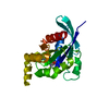
 Basic information
 Components
 Keywords
 TRANSFERASE /
  Function and homology information
 Authors
 Citation
 Structure visualization
 Downloads & links
 Other downloads


 PDBj
PDBj


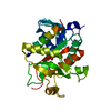
 Assembly
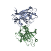


 Mass: 18.015 Da / Num. of mol.: 143 / Source method: isolated from a natural source / Formula: H2O
 Sample preparation
 Processing