 Movie
Movie Controller

[English] 日本語
 Yorodumi
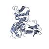
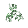
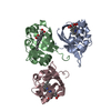
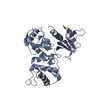
- PDB-3ckd: Crystal structure of the C-terminal domain of the Shigella type I... -
+ Open data

- Basic information
| Entry | Database: PDB / ID: 3ckd | ||||||
|---|---|---|---|---|---|---|---|
| Title | Crystal structure of the C-terminal domain of the Shigella type III effector IpaH | ||||||
 Components Components | Invasion plasmid antigen, secreted by the Mxi-Spa secretion machinery | ||||||
 Keywords Keywords |  LIGASE / E3 ubiquitin ligase / helical / type III effector / Structural Genomics / PSI-2 / Protein Structure Initiative / Midwest Center for Structural Genomics / MCSG LIGASE / E3 ubiquitin ligase / helical / type III effector / Structural Genomics / PSI-2 / Protein Structure Initiative / Midwest Center for Structural Genomics / MCSG | ||||||
| Function / homology |  Function and homology information Function and homology informationsymbiont-mediated suppression of host programmed cell death / effector-mediated activation of programmed cell death in host / : / RING-type E3 ubiquitin transferase / ubiquitin-protein transferase activity / ubiquitin protein ligase activity / ubiquitin-dependent protein catabolic process / host cell cytoplasm / protein ubiquitination / extracellular regionSimilarity search - Function | ||||||
| Biological species |  Shigella flexneri 2a str. 301 (bacteria) Shigella flexneri 2a str. 301 (bacteria) | ||||||
| Method | X-RAY DIFFRACTION / SYNCHROTRON / SAD / Resolution: 2.65 Å | ||||||
 Authors Authors | Lam, R. / Singer, A.U. / Cuff, M.E. / Skarina, T. / Kagan, O. / DiLeo, R. / Edwards, A.M. / Joachimiak, A. / Savchenko, A. / Midwest Center for Structural Genomics (MCSG) | ||||||
 Citation Citation | Journal: Nat.Struct.Mol.Biol. / Year: 2008 Title: Structure of the Shigella T3SS effector IpaH defines a new class of E3 ubiquitin ligases. Authors: Singer, A.U. / Rohde, J.R. / Lam, R. / Skarina, T. / Kagan, O. / Dileo, R. / Chirgadze, N.Y. / Cuff, M.E. / Joachimiak, A. / Tyers, M. / Sansonetti, P.J. / Parsot, C. / Savchenko, A. | ||||||
| History |
|
- Structure visualization
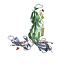
| Structure viewer | Molecule: MolmilJmol/JSmol |
|---|
- Downloads & links
-Download
| PDBx/mmCIF format | 3ckd.cif.gz | 161.4 KB | Display | PDBx/mmCIF format |
|---|---|---|---|---|
| PDB format | pdb3ckd.ent.gz | 135.5 KB | Display | PDB format |
| PDBx/mmJSON format | 3ckd.json.gz | Tree view | PDBx/mmJSON format | |
| Others |  Other downloads Other downloads |
-Validation report
| Arichive directory | https://data.pdbj.org/pub/pdb/validation_reports/ck/3ckdftp://data.pdbj.org/pub/pdb/validation_reports/ck/3ckd | HTTPS FTP |
|---|
-Related structure data
| Similar structure data | |
|---|---|
| Other databases |


-Links
 PDBj
PDBj


- Assembly
| Deposited unit | 
| ||||||||
|---|---|---|---|---|---|---|---|---|---|
| 1 | 
| ||||||||
| 2 | 
| ||||||||
| 3 | 
| ||||||||
| Unit cell |
|
-Components
| #1: Protein | Mass: 35641.309 Da / Num. of mol.: 3 / Fragment: C-terminal ligase domain: Residues 265-575 Source method: isolated from a genetically manipulated source Source: (gene. exp.) Shigella flexneri 2a str. 301 (bacteria)Species: Shigella flexneri / Strain: 301 / Serotype 2a / Gene: ipaH1.4, CP0265 / Plasmid: p15-TV-LIC / Production host: Escherichia coli (E. coli) / Strain (production host): BL21-Gold(DE3)References: UniProt: Q8VSA1, UniProt: P18014*PLUS, ubiquitin-protein ligase#2: Chemical | Sulfate  Mass: 96.063 Da / Num. of mol.: 2 / Source method: obtained synthetically / Formula: SO4 Mass: 96.063 Da / Num. of mol.: 2 / Source method: obtained synthetically / Formula: SO4#3: Chemical | Glycerol  Mass: 92.094 Da / Num. of mol.: 3 / Source method: obtained synthetically / Formula: C3H8O3 Mass: 92.094 Da / Num. of mol.: 3 / Source method: obtained synthetically / Formula: C3H8O3#4: Chemical | ChemComp-PEG / | Diethylene glycol  Mass: 106.120 Da / Num. of mol.: 1 / Source method: obtained synthetically / Formula: C4H10O3 Mass: 106.120 Da / Num. of mol.: 1 / Source method: obtained synthetically / Formula: C4H10O3#5: Water | ChemComp-HOH / | Water Mass: 18.015 Da / Num. of mol.: 24 / Source method: isolated from a natural source / Formula: H2O Mass: 18.015 Da / Num. of mol.: 24 / Source method: isolated from a natural source / Formula: H2O |
|---|
-Experimental details
-Experiment
| Experiment | Method: X-RAY DIFFRACTION / Number of used crystals: 1 |
|---|
- Sample preparation
| Crystal | Density Matthews: 2.75 Å3/Da / Density % sol: 55.24 % |
|---|---|
| Crystal grow | Temperature: 298 K / Method: vapor diffusion, sitting drop Details: 2M Ammonium sulfate, 1% PEG MME 2000, VAPOR DIFFUSION, SITTING DROP, temperature 298K |
-Data collection
| Diffraction | Mean temperature: 100 K |
|---|---|
| Diffraction source | Source: SYNCHROTRON / Site: APS  / Beamline: 19-ID / Wavelength: 0.97845 Å / Beamline: 19-ID / Wavelength: 0.97845 Å |
| Detector | Type: ADSC QUANTUM 315 / Detector: CCD / Date: Oct 7, 2006 / Details: Mirrors |
| Radiation | Monochromator: Double crystal / Protocol: SINGLE WAVELENGTH / Monochromatic (M) / Laue (L): M / Scattering type: x-ray |
| Radiation wavelength | Wavelength: 0.97845 Å / Relative weight: 1 |
| Reflection | Resolution: 2.65→50 Å / Num. all: 35079 / Num. obs: 34738 / % possible obs: 99 % / Observed criterion σ(F): 1 / Observed criterion σ(I): 1 / Redundancy: 8.2 % / Rmerge(I) obs: 0.073 / Net I/σ(I): 35.3 |
| Reflection shell | Resolution: 2.65→2.74 Å / Redundancy: 8.1 % / Rmerge(I) obs: 0.479 / Mean I/σ(I) obs: 3.8 / Num. unique all: 3447 / % possible all: 99.5 |
- Processing
| Software |
| ||||||||||||||||||||||||||||||||||||||||||||||||||||||||||||||||||||||||||||||||||||||||||||||||||||||||||||||||||||||||||||||||||||||||||||||||||||||||||||||||||||||||||
|---|---|---|---|---|---|---|---|---|---|---|---|---|---|---|---|---|---|---|---|---|---|---|---|---|---|---|---|---|---|---|---|---|---|---|---|---|---|---|---|---|---|---|---|---|---|---|---|---|---|---|---|---|---|---|---|---|---|---|---|---|---|---|---|---|---|---|---|---|---|---|---|---|---|---|---|---|---|---|---|---|---|---|---|---|---|---|---|---|---|---|---|---|---|---|---|---|---|---|---|---|---|---|---|---|---|---|---|---|---|---|---|---|---|---|---|---|---|---|---|---|---|---|---|---|---|---|---|---|---|---|---|---|---|---|---|---|---|---|---|---|---|---|---|---|---|---|---|---|---|---|---|---|---|---|---|---|---|---|---|---|---|---|---|---|---|---|---|---|---|---|---|
| Refinement | Method to determine structure: SAD / Resolution: 2.65→49.21 Å / Cor.coef. Fo:Fc: 0.928 / Cor.coef. Fo:Fc free: 0.891 / SU B: 22.903 / SU ML: 0.234 / Cross valid method: THROUGHOUT / σ(F): 0 / ESU R: 0.561 / ESU R Free: 0.336 / Stereochemistry target values: MAXIMUM LIKELIHOOD / Details: HYDROGENS HAVE BEEN ADDED IN THE RIDING POSITIONS
| ||||||||||||||||||||||||||||||||||||||||||||||||||||||||||||||||||||||||||||||||||||||||||||||||||||||||||||||||||||||||||||||||||||||||||||||||||||||||||||||||||||||||||
| Solvent computation | Ion probe radii: 0.8 Å / Shrinkage radii: 0.8 Å / VDW probe radii: 1.2 Å / Solvent model: BABINET MODEL WITH MASK | ||||||||||||||||||||||||||||||||||||||||||||||||||||||||||||||||||||||||||||||||||||||||||||||||||||||||||||||||||||||||||||||||||||||||||||||||||||||||||||||||||||||||||
| Displacement parameters | Biso mean: 61.151 Å2
| ||||||||||||||||||||||||||||||||||||||||||||||||||||||||||||||||||||||||||||||||||||||||||||||||||||||||||||||||||||||||||||||||||||||||||||||||||||||||||||||||||||||||||
| Refinement step | Cycle: LAST / Resolution: 2.65→49.21 Å
| ||||||||||||||||||||||||||||||||||||||||||||||||||||||||||||||||||||||||||||||||||||||||||||||||||||||||||||||||||||||||||||||||||||||||||||||||||||||||||||||||||||||||||
| Refine LS restraints |
| ||||||||||||||||||||||||||||||||||||||||||||||||||||||||||||||||||||||||||||||||||||||||||||||||||||||||||||||||||||||||||||||||||||||||||||||||||||||||||||||||||||||||||
| LS refinement shell | Resolution: 2.65→2.719 Å / Total num. of bins used: 20
|
