 Movie
Movie Controller


+ Open data

- Basic information
| Entry | Database: PDB / ID: 3chk | ||||||
|---|---|---|---|---|---|---|---|
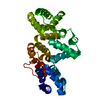
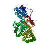
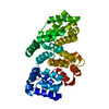
| Title | Calcium bound structure of alpha-14 giardin | ||||||
 Components Components | Alpha-14 giardin | ||||||
 Keywords Keywords | METAL BINDING PROTEIN / Calcium-binding /  Annexin Annexin | ||||||
| Function / homology |  Function and homology information Function and homology information1-phosphatidylinositol binding / cell projection organization / phosphatidylethanolamine binding / calcium-dependent phospholipid binding / motile cilium / phosphatidylserine binding / heparin binding / calcium ion binding / nucleus / plasma membrane / cytoplasmSimilarity search - Function | ||||||
| Biological species |  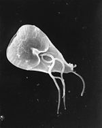 Giardia lamblia (eukaryote) Giardia lamblia (eukaryote) | ||||||
| Method | X-RAY DIFFRACTION / SYNCHROTRON / Resolution: 1.65 Å | ||||||
 Authors Authors | Pathuri, P. / Luecke, H. | ||||||
 Citation Citation | Journal: J.Mol.Biol. / Year: 2009 Title: Apo and calcium-bound crystal structures of cytoskeletal protein alpha-14 giardin (annexin E1) from the intestinal protozoan parasite Giardia lamblia Authors: Pathuri, P. / Nguyen, E.T. / Ozorowski, G. / Svard, S.G. / Luecke, H. | ||||||
| History |
|
- Structure visualization
| Structure viewer | Molecule: MolmilJmol/JSmol |
|---|
- Downloads & links
-Download
| PDBx/mmCIF format | 3chk.cif.gz | 83.6 KB | Display | PDBx/mmCIF format |
|---|---|---|---|---|
| PDB format | pdb3chk.ent.gz | 61.9 KB | Display | PDB format |
| PDBx/mmJSON format | 3chk.json.gz | Tree view | PDBx/mmJSON format | |
| Others |  Other downloads Other downloads |
-Validation report
| Arichive directory | https://data.pdbj.org/pub/pdb/validation_reports/ch/3chkftp://data.pdbj.org/pub/pdb/validation_reports/ch/3chk | HTTPS FTP |
|---|
-Related structure data


-Links
 PDBj
PDBj- Assembly
| Deposited unit | 
| ||||||||
|---|---|---|---|---|---|---|---|---|---|
| 1 |
| ||||||||
| Unit cell |
|
-Components
| #1: Protein | Mass: 38637.184 Da / Num. of mol.: 1 Source method: isolated from a genetically manipulated source Source: (gene. exp.) Giardia lamblia (eukaryote) / Gene: Alpha-14 / Production host:  Escherichia coli (E. coli) / References: UniProt: Q4VPP4, UniProt: Q9NFS4*PLUS Escherichia coli (E. coli) / References: UniProt: Q4VPP4, UniProt: Q9NFS4*PLUS | ||
|---|---|---|---|
| #2: Chemical | ChemComp-CA /   Mass: 40.078 Da / Num. of mol.: 5 / Source method: obtained synthetically / Formula: Ca Mass: 40.078 Da / Num. of mol.: 5 / Source method: obtained synthetically / Formula: Ca#3: Water | ChemComp-HOH / | Water Mass: 18.015 Da / Num. of mol.: 367 / Source method: isolated from a natural source / Formula: H2O Mass: 18.015 Da / Num. of mol.: 367 / Source method: isolated from a natural source / Formula: H2O |
-Experimental details
-Experiment
| Experiment | Method: X-RAY DIFFRACTION / Number of used crystals: 1 |
|---|
- Sample preparation
| Crystal | Density Matthews: 2.15 Å3/Da / Density % sol: 42.87 % |
|---|---|
| Crystal grow | Temperature: 298 K / Method: vapor diffusion, sitting drop / pH: 6.5 Details: 50 mM calcium chloride, 100 mM bis-Tris pH 6.5, and 24% (w/v) PEG 4000, VAPOR DIFFUSION, SITTING DROP, temperature 298K |
-Data collection
| Diffraction source | Source: SYNCHROTRON / Site: SSRL  / Beamline: BL11-1 / Wavelength: 0.979 / Beamline: BL11-1 / Wavelength: 0.979 |
|---|---|
| Detector | Type: MARMOSAIC 325 mm CCD / Detector: CCD / Date: Apr 23, 2007 |
| Radiation | Protocol: SINGLE WAVELENGTH / Monochromatic (M) / Laue (L): M / Scattering type: x-ray |
| Radiation wavelength | Wavelength: 0.979 Å / Relative weight: 1 |
| Reflection | Resolution: 1.65→50 Å / Num. all: 38460 / Num. obs: 38460 / % possible obs: 100 % / Observed criterion σ(F): 0 / Observed criterion σ(I): 0 |
- Processing
| Software |
| |||||||||||||||||||||||||||||||||||||||||||||||||||||||||||||||||||||||||||||||||||||||||||||||
|---|---|---|---|---|---|---|---|---|---|---|---|---|---|---|---|---|---|---|---|---|---|---|---|---|---|---|---|---|---|---|---|---|---|---|---|---|---|---|---|---|---|---|---|---|---|---|---|---|---|---|---|---|---|---|---|---|---|---|---|---|---|---|---|---|---|---|---|---|---|---|---|---|---|---|---|---|---|---|---|---|---|---|---|---|---|---|---|---|---|---|---|---|---|---|---|---|
| Refinement | Resolution: 1.65→50 Å / Cor.coef. Fo:Fc: 0.958 / Cor.coef. Fo:Fc free: 0.941 / SU B: 2.385 / SU ML: 0.082 / Cross valid method: THROUGHOUT / σ(F): 0 / ESU R: 0.114 / ESU R Free: 0.112 / Stereochemistry target values: MAXIMUM LIKELIHOOD / Details: HYDROGENS HAVE BEEN ADDED IN THE RIDING POSITIONS
| |||||||||||||||||||||||||||||||||||||||||||||||||||||||||||||||||||||||||||||||||||||||||||||||
| Solvent computation | Ion probe radii: 0.8 Å / Shrinkage radii: 0.8 Å / VDW probe radii: 1.2 Å / Solvent model: MASK | |||||||||||||||||||||||||||||||||||||||||||||||||||||||||||||||||||||||||||||||||||||||||||||||
| Displacement parameters | Biso mean: 27.172 Å2
| |||||||||||||||||||||||||||||||||||||||||||||||||||||||||||||||||||||||||||||||||||||||||||||||
| Refinement step | Cycle: LAST / Resolution: 1.65→50 Å
| |||||||||||||||||||||||||||||||||||||||||||||||||||||||||||||||||||||||||||||||||||||||||||||||
| Refine LS restraints |
| |||||||||||||||||||||||||||||||||||||||||||||||||||||||||||||||||||||||||||||||||||||||||||||||
| LS refinement shell | Resolution: 1.65→1.694 Å / Total num. of bins used: 20
|
