 Movie
Movie Controller

[English] 日本語
 Yorodumi
- PDB-3cgo: IRAK-4 Inhibitors (Part II)- A structure based assessment of imid... -
+ Open data

- Basic information
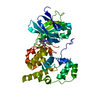
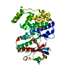
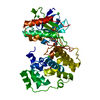
| Entry | Database: PDB / ID: 3cgo | ||||||
|---|---|---|---|---|---|---|---|
| Title | IRAK-4 Inhibitors (Part II)- A structure based assessment of imidazo[1,2 a]pyridine binding | ||||||
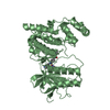
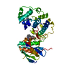
 Components Components | Mitogen-activated protein kinase 10 | ||||||
 Keywords Keywords |  TRANSFERASE / JNK3 kinase / Inhibitor / Alternative splicing / ATP-binding / Chromosomal rearrangement / Cytoplasm / Epilepsy / Nucleotide-binding / Phosphoprotein / Serine/threonine-protein kinase TRANSFERASE / JNK3 kinase / Inhibitor / Alternative splicing / ATP-binding / Chromosomal rearrangement / Cytoplasm / Epilepsy / Nucleotide-binding / Phosphoprotein / Serine/threonine-protein kinase | ||||||
| Function / homology |  Function and homology information Function and homology informationJUN kinase activity / Activation of the AP-1 family of transcription factors / Fc-epsilon receptor signaling pathway / MAP kinase kinase activity / response to light stimulus / mitogen-activated protein kinase / JNK cascade / JNK (c-Jun kinases) phosphorylation and activation mediated by activated human TAK1 / FCERI mediated MAPK activation / regulation of circadian rhythm ...JUN kinase activity / Activation of the AP-1 family of transcription factors / Fc-epsilon receptor signaling pathway / MAP kinase kinase activity / response to light stimulus / mitogen-activated protein kinase / JNK cascade / JNK (c-Jun kinases) phosphorylation and activation mediated by activated human TAK1 / FCERI mediated MAPK activation / regulation of circadian rhythm / cellular senescence / rhythmic process / Oxidative Stress Induced Senescence / protein phosphorylation / protein serine kinase activity / signal transduction / mitochondrion / nucleoplasm / ATP binding / nucleus / plasma membrane / cytosol / cytoplasmSimilarity search - Function | ||||||
| Biological species |  Homo sapiens (human) Homo sapiens (human) | ||||||
| Method | X-RAY DIFFRACTION / MOLECULAR REPLACEMENT / Resolution: 3 Å | ||||||
 Authors Authors | Ceska, T.A. / Platt, A. / Fortunato, M. / Dickson, K.M. / Beevers, R. | ||||||
 Citation Citation | Journal: Bioorg.Med.Chem.Lett. / Year: 2008 Title: IRAK-4 inhibitors. Part II: A structure-based assessment of imidazo[1,2-a]pyridine binding Authors: Buckley, G.M. / Ceska, T.A. / Fraser, J.L. / Gowers, L. / Groom, C.R. / Higueruelo, A.P. / Jenkins, K. / Mack, S.R. / Morgan, T. / Parry, D.M. / Pitt, W.R. / Rausch, O. / Richard, M.D. / Sabin, V. | ||||||
| History |
|
- Structure visualization
| Structure viewer | Molecule: MolmilJmol/JSmol |
|---|
- Downloads & links
-Download
| PDBx/mmCIF format | 3cgo.cif.gz | 83.3 KB | Display | PDBx/mmCIF format |
|---|---|---|---|---|
| PDB format | pdb3cgo.ent.gz | 61.1 KB | Display | PDB format |
| PDBx/mmJSON format | 3cgo.json.gz | Tree view | PDBx/mmJSON format | |
| Others |  Other downloads Other downloads |
-Validation report
| Arichive directory | https://data.pdbj.org/pub/pdb/validation_reports/cg/3cgoftp://data.pdbj.org/pub/pdb/validation_reports/cg/3cgo | HTTPS FTP |
|---|
-Related structure data
| Related structure data |  3cgfC  1jnkS C: citing same article ( S: Starting model for refinement |
|---|---|
| Similar structure data |


-Links
 PDBj
PDBj


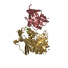
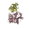
- Assembly
| Deposited unit | 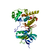
| ||||||||
|---|---|---|---|---|---|---|---|---|---|
| 1 |
| ||||||||
| Unit cell |
|
-Components
| #1: Protein | Mass: 42116.727 Da / Num. of mol.: 1 / Fragment: Protein kinase domain, UNP residues 40-402 Source method: isolated from a genetically manipulated source Source: (gene. exp.) Homo sapiens (human) / Gene: MAPK10, JNK3, JNK3A, PRKM10 / Production host:  Escherichia coli (E. coli) Escherichia coli (E. coli)References: UniProt: P53779, mitogen-activated protein kinase |
|---|---|
| #2: Chemical | ChemComp-JNO /   Mass: 365.432 Da / Num. of mol.: 1 / Source method: obtained synthetically / Formula: C19H23N7O Mass: 365.432 Da / Num. of mol.: 1 / Source method: obtained synthetically / Formula: C19H23N7O |
| #3: Water | ChemComp-HOH / Water Mass: 18.015 Da / Num. of mol.: 54 / Source method: isolated from a natural source / Formula: H2O Mass: 18.015 Da / Num. of mol.: 54 / Source method: isolated from a natural source / Formula: H2O |
-Experimental details
-Experiment
| Experiment | Method: X-RAY DIFFRACTION / Number of used crystals: 1 |
|---|
- Sample preparation
| Crystal | Density Matthews: 2.36 Å3/Da / Density % sol: 47.9 % |
|---|---|
| Crystal grow | pH: 7 / Details: pH7.0 |
-Data collection
| Diffraction | Mean temperature: 100 K |
|---|---|
| Diffraction source | Source: ROTATING ANODE / Type: RIGAKU RU300 / Wavelength: 1.5418 / Wavelength: 1.5418 Å |
| Detector | Type: MAR scanner 345 mm plate / Detector: IMAGE PLATE / Date: Aug 27, 2004 / Details: OSMIC |
| Radiation | Protocol: SINGLE WAVELENGTH / Monochromatic (M) / Laue (L): M / Scattering type: x-ray |
| Radiation wavelength | Wavelength: 1.5418 Å / Relative weight: 1 |
| Reflection | Resolution: 2.5→20 Å / Num. obs: 13086 / % possible obs: 88.4 % / Observed criterion σ(I): 0 / Redundancy: 8.89 % / Rmerge(I) obs: 0.098 / Net I/σ(I): 11.6 |
| Reflection shell | Resolution: 2.5→2.54 Å / Rmerge(I) obs: 0.498 / Mean I/σ(I) obs: 1.3 / % possible all: 59.1 |
- Processing
| Software |
| ||||||||||||||||||||||||||||||||||||||||||||||||||||||||||||
|---|---|---|---|---|---|---|---|---|---|---|---|---|---|---|---|---|---|---|---|---|---|---|---|---|---|---|---|---|---|---|---|---|---|---|---|---|---|---|---|---|---|---|---|---|---|---|---|---|---|---|---|---|---|---|---|---|---|---|---|---|---|
| Refinement | Method to determine structure: MOLECULAR REPLACEMENT Starting model: PDB ENTRY 1JNK Resolution: 3→20 Å / Rfactor Rfree error: 0.008 / Data cutoff high absF: 99999 / Data cutoff low absF: 0 / Isotropic thermal model: RESTRAINED / Cross valid method: THROUGHOUT / σ(F): 0
| ||||||||||||||||||||||||||||||||||||||||||||||||||||||||||||
| Solvent computation | Solvent model: FLAT MODEL / Bsol: 27.1 Å2 / ksol: 0.28 e/Å3 | ||||||||||||||||||||||||||||||||||||||||||||||||||||||||||||
| Refinement step | Cycle: LAST / Resolution: 3→20 Å
| ||||||||||||||||||||||||||||||||||||||||||||||||||||||||||||
| Refine LS restraints |
| ||||||||||||||||||||||||||||||||||||||||||||||||||||||||||||
| Xplor file |
|
