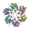
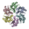
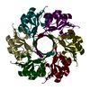
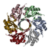
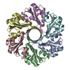
A: Propanediol utilization protein pduU B: Propanediol utilization protein pduU C: Propanediol utilization protein pduU D: Propanediol utilization protein pduU
A: Propanediol utilization protein pduU B: Propanediol utilization protein pduU C: Propanediol utilization protein pduU D: Propanediol utilization protein pduU
A: Propanediol utilization protein pduU B: Propanediol utilization protein pduU C: Propanediol utilization protein pduU D: Propanediol utilization protein pduU
A: Propanediol utilization protein pduU B: Propanediol utilization protein pduU C: Propanediol utilization protein pduU D: Propanediol utilization protein pduU
Resolution: 1.8→61.59 Å / Cor.coef. Fo:Fc: 0.953 / Cor.coef. Fo:Fc free: 0.94 / SU B: 7.761 / SU ML: 0.116 / TLS residual ADP flag: LIKELY RESIDUAL / Cross valid method: THROUGHOUT / σ(F): 0 / ESU R: 0.146 / ESU R Free: 0.137 / Stereochemistry target values: MAXIMUM LIKELIHOOD Details: Hydrogens have been added in the riding positions. Residue Asp41 from chains A, B, C, and D have an unusual torsion angles due to crystallographic packing.
Rfactor
Num. reflection
% reflection
Selection details
Rfree
0.242
2063
5 %
RANDOM
Rwork
0.203
-
-
-
obs
0.205
40944
99.09 %
-
Solvent computation
Ion probe radii: 0.8 Å / Shrinkage radii: 0.8 Å / VDW probe radii: 1.4 Å / Solvent model: MASK
Displacement parameters
Biso mean: 23.646 Å2
Baniso -1
Baniso -2
Baniso -3
1-
-2.55 Å2
-1.27 Å2
0 Å2
2-
-
-2.55 Å2
0 Å2
3-
-
-
3.82 Å2
Refine analyze
Free
Obs
Luzzati coordinate error
0.137 Å
0.146 Å
Refinement step
Cycle: LAST / Resolution: 1.8→61.59 Å
Protein
Nucleic acid
Ligand
Solvent
Total
Num. atoms
3458
0
0
165
3623
Refine LS restraints
Refine-ID
Type
Dev ideal
Dev ideal target
Number
X-RAY DIFFRACTION
r_bond_refined_d
0.012
0.022
3583
X-RAY DIFFRACTION
r_bond_other_d
0.001
0.02
2343
X-RAY DIFFRACTION
r_angle_refined_deg
1.431
1.97
4873
X-RAY DIFFRACTION
r_angle_other_deg
0.905
3
5807
X-RAY DIFFRACTION
r_dihedral_angle_1_deg
5.413
5
478
X-RAY DIFFRACTION
r_dihedral_angle_2_deg
39.27
24.815
135
X-RAY DIFFRACTION
r_dihedral_angle_3_deg
11.858
15
646
X-RAY DIFFRACTION
r_dihedral_angle_4_deg
10.075
15
17
X-RAY DIFFRACTION
r_chiral_restr
0.085
0.2
610
X-RAY DIFFRACTION
r_gen_planes_refined
0.004
0.02
3950
X-RAY DIFFRACTION
r_gen_planes_other
0.001
0.02
659
X-RAY DIFFRACTION
r_nbd_refined
0.197
0.2
668
X-RAY DIFFRACTION
r_nbd_other
0.183
0.2
2296
X-RAY DIFFRACTION
r_nbtor_refined
0.171
0.2
1753
X-RAY DIFFRACTION
r_nbtor_other
0.086
0.2
1897
X-RAY DIFFRACTION
r_xyhbond_nbd_refined
0.154
0.2
144
X-RAY DIFFRACTION
r_symmetry_vdw_refined
0.192
0.2
24
X-RAY DIFFRACTION
r_symmetry_vdw_other
0.297
0.2
89
X-RAY DIFFRACTION
r_symmetry_hbond_refined
0.206
0.2
18
X-RAY DIFFRACTION
r_mcbond_it
2.657
2
3039
X-RAY DIFFRACTION
r_mcbond_other
0.714
2
951
X-RAY DIFFRACTION
r_mcangle_it
3.012
3
3756
X-RAY DIFFRACTION
r_scbond_it
2.433
2
1409
X-RAY DIFFRACTION
r_scangle_it
3.334
3
1107
LS refinement shell
Resolution: 1.8→1.848 Å / Total num. of bins used: 20
Rfactor
Num. reflection
% reflection
Rfree
0.31
141
-
Rwork
0.292
2638
-
all
-
2779
-
obs
-
-
91.38 %
Refinement TLS params.
Method: refined / Refine-ID: X-RAY DIFFRACTION
ID
L11 (°2)
L12 (°2)
L13 (°2)
L22 (°2)
L23 (°2)
L33 (°2)
S11 (Å °)
S12 (Å °)
S13 (Å °)
S21 (Å °)
S22 (Å °)
S23 (Å °)
S31 (Å °)
S32 (Å °)
S33 (Å °)
T11 (Å2)
T12 (Å2)
T13 (Å2)
T22 (Å2)
T23 (Å2)
T33 (Å2)
Origin x (Å)
Origin y (Å)
Origin z (Å)
1
0.7358
-0.0594
-0.5675
0.2339
-0.1697
0.7053
0.0048
-0.0301
0.0138
0.0279
-0.0245
-0.0508
-0.0146
0.0528
0.0197
-0.0251
0.0069
-0.0067
-0.0019
-0.009
-0.0212
19.9459
1.2597
15.2468
2
0.9275
0.418
-0.2017
0.5347
-0.4428
0.5719
-0.0041
-0.0182
0.1623
0.0227
0.0096
0.0206
-0.0117
0.0154
-0.0056
-0.0123
-0.0071
-0.0016
-0.0213
-0.0058
-0.025
8.913
17.9421
15.2975
3
0.4157
0.066
0.1925
0.9224
0.2657
0.6297
0.0281
0.0122
0.0257
-0.064
-0.0255
-0.1989
-0.0578
0.0088
-0.0026
-0.0076
-0.0191
0.0059
-0.0109
0.011
-0.0467
15.8906
11.4877
-22.8582
4
1.113
0.0326
0.401
0.3145
-0.0541
0.6731
0.0236
0.0284
-0.11
-0.0356
-0.0159
-0.1021
0.045
0.0624
-0.0077
-0.0124
0.0032
0.0142
0.02
0.0124
-0.0769
18.3615
-8.4882
-22.3922
Refinement TLS group
ID
Refine-ID
Refine TLS-ID
Auth asym-ID
Label asym-ID
Auth seq-ID
Label seq-ID
1
X-RAY DIFFRACTION
1
A
A
7 - 118
7 - 118
2
X-RAY DIFFRACTION
2
B
B
7 - 118
7 - 118
3
X-RAY DIFFRACTION
3
C
C
4 - 122
4 - 122
4
X-RAY DIFFRACTION
4
D
D
6 - 122
6 - 122
+
About Yorodumi
-
News
-
Feb 9, 2022. New format data for meta-information of EMDB entries
New format data for meta-information of EMDB entries
Version 3 of the EMDB header file is now the official format.
The previous official version 1.9 will be removed from the archive.
In the structure databanks used in Yorodumi, some data are registered as the other names, "COVID-19 virus" and "2019-nCoV". Here are the details of the virus and the list of structure data.
Jan 31, 2019. EMDB accession codes are about to change! (news from PDBe EMDB page)
EMDB accession codes are about to change! (news from PDBe EMDB page)
The allocation of 4 digits for EMDB accession codes will soon come to an end. Whilst these codes will remain in use, new EMDB accession codes will include an additional digit and will expand incrementally as the available range of codes is exhausted. The current 4-digit format prefixed with “EMD-” (i.e. EMD-XXXX) will advance to a 5-digit format (i.e. EMD-XXXXX), and so on. It is currently estimated that the 4-digit codes will be depleted around Spring 2019, at which point the 5-digit format will come into force.
The EM Navigator/Yorodumi systems omit the EMD- prefix.
Related info.:Q: What is EMD? / ID/Accession-code notation in Yorodumi/EM Navigator
Yorodumi is a browser for structure data from EMDB, PDB, SASBDB, etc.
This page is also the successor to EM Navigator detail page, and also detail information page/front-end page for Omokage search.
The word "yorodu" (or yorozu) is an old Japanese word meaning "ten thousand". "mi" (miru) is to see.
Related info.:EMDB / PDB / SASBDB / Comparison of 3 databanks / Yorodumi Search / Aug 31, 2016. New EM Navigator & Yorodumi / Yorodumi Papers / Jmol/JSmol / Function and homology information / Changes in new EM Navigator and Yorodumi
 Movie
Movie Controller

 Yorodumi
 Open data
 Basic information
 Components
 Keywords
 Function and homology information
 Salmonella typhimurium (bacteria)
 X-RAY DIFFRACTION /
  Authors
 Citation
 Structure visualization
 Downloads & links
 Other downloads
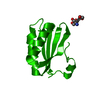


 PDBj
PDBj


 Assembly


 Mass: 18.015 Da / Num. of mol.: 165 / Source method: isolated from a natural source / Formula: H2O
 Sample preparation
 / Beamline: 8.2.2 / Wavelength: 1 Å
 Processing