 Movie
Movie Controller


+ Open data

- Basic information
| Entry | Database: PDB / ID: 3cfw | |||||||||
|---|---|---|---|---|---|---|---|---|---|---|
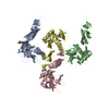
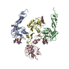
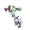
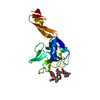
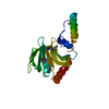
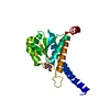
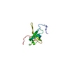
| Title | L-selectin lectin and EGF domains | |||||||||
 Components Components | L-selectin | |||||||||
 Keywords Keywords | CELL ADHESION / L-selectin / lectin / EGF / EGF-like domain / Glycoprotein / Membrane / Sushi / Transmembrane | |||||||||
| Function / homology |  Function and homology informationglycosphingolipid binding / sialic acid binding / oligosaccharide binding / leukocyte tethering or rolling / calcium-dependent cell-cell adhesion via plasma membrane cell adhesion molecules / heterophilic cell-cell adhesion via plasma membrane cell adhesion molecules / leukocyte cell-cell adhesion / secretory granule membrane / response to cytokine / Cell surface interactions at the vascular wall ...glycosphingolipid binding / sialic acid binding / oligosaccharide binding / leukocyte tethering or rolling / calcium-dependent cell-cell adhesion via plasma membrane cell adhesion molecules / heterophilic cell-cell adhesion via plasma membrane cell adhesion molecules / leukocyte cell-cell adhesion / secretory granule membrane / response to cytokine / Cell surface interactions at the vascular wall / Immunoregulatory interactions between a Lymphoid and a non-Lymphoid cell / heparin binding / carbohydrate binding / protease binding / cell adhesion / external side of plasma membrane / calcium ion binding / Neutrophil degranulation / extracellular space / plasma membrane Function and homology informationglycosphingolipid binding / sialic acid binding / oligosaccharide binding / leukocyte tethering or rolling / calcium-dependent cell-cell adhesion via plasma membrane cell adhesion molecules / heterophilic cell-cell adhesion via plasma membrane cell adhesion molecules / leukocyte cell-cell adhesion / secretory granule membrane / response to cytokine / Cell surface interactions at the vascular wall ...glycosphingolipid binding / sialic acid binding / oligosaccharide binding / leukocyte tethering or rolling / calcium-dependent cell-cell adhesion via plasma membrane cell adhesion molecules / heterophilic cell-cell adhesion via plasma membrane cell adhesion molecules / leukocyte cell-cell adhesion / secretory granule membrane / response to cytokine / Cell surface interactions at the vascular wall / Immunoregulatory interactions between a Lymphoid and a non-Lymphoid cell / heparin binding / carbohydrate binding / protease binding / cell adhesion / external side of plasma membrane / calcium ion binding / Neutrophil degranulation / extracellular space / plasma membraneSimilarity search - Function | |||||||||
| Biological species |  Homo sapiens (human) Homo sapiens (human) | |||||||||
| Method | X-RAY DIFFRACTION / SYNCHROTRON / MOLECULAR REPLACEMENT / Resolution: 2.2 Å | |||||||||
 Authors Authors | Mehta, P. / Oganesyan, V. / Terzyan, S. / Mather, T. / McEver, R.P. | |||||||||
 Citation Citation | Journal: J.Biol.Chem. / Year: 2017 Title: Glycan Bound to the Selectin Low Affinity State Engages Glu-88 to Stabilize the High Affinity State under Force. Authors: Mehta-D'souza, P. / Klopocki, A.G. / Oganesyan, V. / Terzyan, S. / Mather, T. / Li, Z. / Panicker, S.R. / Zhu, C. / McEver, R.P. | |||||||||
| History |
|
- Structure visualization
| Structure viewer | Molecule: MolmilJmol/JSmol |
|---|
- Downloads & links
-Download
| PDBx/mmCIF format | 3cfw.cif.gz | 53.6 KB | Display | PDBx/mmCIF format |
|---|---|---|---|---|
| PDB format | pdb3cfw.ent.gz | 36.8 KB | Display | PDB format |
| PDBx/mmJSON format | 3cfw.json.gz | Tree view | PDBx/mmJSON format | |
| Others |  Other downloads Other downloads |
-Validation report
| Arichive directory | https://data.pdbj.org/pub/pdb/validation_reports/cf/3cfwftp://data.pdbj.org/pub/pdb/validation_reports/cf/3cfw | HTTPS FTP |
|---|
-Related structure data
| Related structure data | 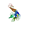 1eslS S: Starting model for refinement |
|---|---|
| Similar structure data |


-Links
 PDBj
PDBj


- Assembly
| Deposited unit | 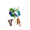
| ||||||||
|---|---|---|---|---|---|---|---|---|---|
| 1 |
| ||||||||
| Unit cell |
|
-Components
| #1: Protein | / Selectin L (Lymphocyte adhesion molecule 1) / isoform CRA_b / Selectin L / Lymphocyte adhesion molecule 1 Mass: 19209.531 Da / Num. of mol.: 1 / Fragment: EGF domain (UNP residues 39-194) Source method: isolated from a genetically manipulated source Source: (gene. exp.) Homo sapiens (human) / Gene: L-selectin, SELL / Plasmid: pEE14.1 / Cell line (production host): Ovary cells / Production host:   Cricetulus griseus (Chinese hamster) / Strain (production host): Lec 1 / References: UniProt: P14151 Cricetulus griseus (Chinese hamster) / Strain (production host): Lec 1 / References: UniProt: P14151 |
|---|---|
| #2: Polysaccharide | alpha-D-mannopyranose-(1-3)-[beta-D-mannopyranose-(1-6)]alpha-D-mannopyranose-(1-4)-2-acetamido-2- ...alpha-D-mannopyranose-(1-3)-[beta-D-mannopyranose-(1-6)]alpha-D-mannopyranose-(1-4)-2-acetamido-2-deoxy-beta-D-glucopyranose-(1-4)-2-acetamido-2-deoxy-beta-D-glucopyranose / Mass: 910.823 Da / Num. of mol.: 1 Source method: isolated from a genetically manipulated source |
| #3: Sugar | ChemComp-NAG / N-Acetylglucosamine  Type: D-saccharide, beta linking / Mass: 221.208 Da / Num. of mol.: 1 Type: D-saccharide, beta linking / Mass: 221.208 Da / Num. of mol.: 1Source method: isolated from a genetically manipulated source Formula: C8H15NO6 |
| #4: Chemical | ChemComp-CA /   Mass: 40.078 Da / Num. of mol.: 1 / Source method: obtained synthetically / Formula: Ca Mass: 40.078 Da / Num. of mol.: 1 / Source method: obtained synthetically / Formula: Ca |
| #5: Water | ChemComp-HOH / Water Mass: 18.015 Da / Num. of mol.: 138 / Source method: isolated from a natural source / Formula: H2O Mass: 18.015 Da / Num. of mol.: 138 / Source method: isolated from a natural source / Formula: H2O |
-Experimental details
-Experiment
| Experiment | Method: X-RAY DIFFRACTION / Number of used crystals: 1 |
|---|
- Sample preparation
| Crystal | Density Matthews: 3.8 Å3/Da / Density % sol: 67.61 % |
|---|---|
| Crystal grow | Temperature: 293 K / Method: vapor diffusion, hanging drop / pH: 8.5 Details: 0.17 M Sodium acetate trihydrate, 0.085 M Tris-HCl, pH 8.5, 25.5 % w/v PEG 4000,15% v/v Glycerol, VAPOR DIFFUSION, HANGING DROP, temperature 293K |
-Data collection
| Diffraction | Mean temperature: 100 K |
|---|---|
| Diffraction source | Source: SYNCHROTRON / Site: NSLS  / Beamline: X12B / Wavelength: 0.9767 Å / Beamline: X12B / Wavelength: 0.9767 Å |
| Detector | Type: ADSC QUANTUM 4 / Detector: CCD / Date: Oct 17, 2000 / Details: mirrors/monochromator |
| Radiation | Monochromator: Si 111 CHANNEL / Protocol: SINGLE WAVELENGTH / Monochromatic (M) / Laue (L): M / Scattering type: x-ray |
| Radiation wavelength | Wavelength: 0.9767 Å / Relative weight: 1 |
| Reflection | Resolution: 2.2→25 Å / Num. all: 14196 / Num. obs: 14192 / % possible obs: 100 % / Observed criterion σ(I): -3 / Redundancy: 5.25 % / Biso Wilson estimate: 29.4 Å2 / Rmerge(I) obs: 0.089 / Net I/σ(I): 12.8 |
| Reflection shell | Resolution: 2.2→2.28 Å / Rmerge(I) obs: 0.644 / Mean I/σ(I) obs: 2.32 / Num. unique all: 1402 / % possible all: 100 |
- Processing
| Software |
| |||||||||||||||||||||||||
|---|---|---|---|---|---|---|---|---|---|---|---|---|---|---|---|---|---|---|---|---|---|---|---|---|---|---|
| Refinement | Method to determine structure: MOLECULAR REPLACEMENT Starting model: 1ESL Resolution: 2.2→25 Å / Isotropic thermal model: Isotropic / Cross valid method: THROUGHOUT / σ(F): 0 / Stereochemistry target values: Engh & Huber
| |||||||||||||||||||||||||
| Displacement parameters | Biso mean: 34.896 Å2 | |||||||||||||||||||||||||
| Refinement step | Cycle: LAST / Resolution: 2.2→25 Å
| |||||||||||||||||||||||||
| Refine LS restraints |
|
