 Movie
Movie Controller

[English] 日本語
 Yorodumi
- PDB-1esl: INSIGHT INTO E-SELECTIN(SLASH)LIGAND INTERACTION FROM THE CRYSTAL... -
+ Open data

- Basic information
| Entry | Database: PDB / ID: 1esl | ||||||
|---|---|---|---|---|---|---|---|
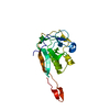
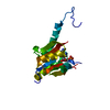
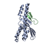
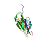
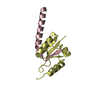
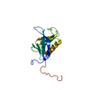
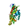
| Title | INSIGHT INTO E-SELECTIN(SLASH)LIGAND INTERACTION FROM THE CRYSTAL STRUCTURE AND MUTAGENESIS OF THE LEC(SLASH)EGF DOMAINS | ||||||
 Components Components | HUMAN E-SELECTIN | ||||||
 Keywords Keywords | CELL ADHESION PROTEIN | ||||||
| Function / homology |  Function and homology information Function and homology informationactin filament-based process / oligosaccharide binding / positive regulation of leukocyte tethering or rolling / sialic acid binding / leukocyte migration involved in inflammatory response / leukocyte tethering or rolling / positive regulation of leukocyte migration / heterophilic cell-cell adhesion via plasma membrane cell adhesion molecules / leukocyte cell-cell adhesion / cortical cytoskeleton ...actin filament-based process / oligosaccharide binding / positive regulation of leukocyte tethering or rolling / sialic acid binding / leukocyte migration involved in inflammatory response / leukocyte tethering or rolling / positive regulation of leukocyte migration / heterophilic cell-cell adhesion via plasma membrane cell adhesion molecules / leukocyte cell-cell adhesion / cortical cytoskeleton / phospholipase binding / positive regulation of receptor internalization / activation of phospholipase C activity / response to tumor necrosis factor / clathrin-coated pit / response to interleukin-1 / response to cytokine / caveola / calcium-mediated signaling / Cell surface interactions at the vascular wall / transmembrane signaling receptor activity / regulation of inflammatory response / response to lipopolysaccharide / inflammatory response / membrane raft / external side of plasma membrane / perinuclear region of cytoplasm / extracellular space / metal ion binding / plasma membraneSimilarity search - Function | ||||||
| Biological species |  Homo sapiens (human) Homo sapiens (human) | ||||||
| Method | X-RAY DIFFRACTION / Resolution: 2 Å | ||||||
 Authors Authors | Graves, B.J. / Crowther, R.L. | ||||||
 Citation Citation | Journal: Nature / Year: 1994 Title: Insight into E-selectin/ligand interaction from the crystal structure and mutagenesis of the lec/EGF domains. Authors: Graves, B.J. / Crowther, R.L. / Chandran, C. / Rumberger, J.M. / Li, S. / Huang, K.S. / Presky, D.H. / Familletti, P.C. / Wolitzky, B.A. / Burns, D.K. #1: Journal: J.Biol.Chem. / Year: 1994Title: Consensus Repeat Domains of E-Selectin Enhance Ligand Binding Authors: Li, S.H. / Burns, D.K. / Rumberger, J.M. / Presky, D.H. / Wilkinson, V.L. / Anostario Junior, M. / Wolitzky, B.A. / Norton, C.R. / Familletti, P.C. / Kim, K.J. / Goldstein, A.L. / Cox, D.C. / Huang, K.-S. | ||||||
| History |
| ||||||
| Remark 650 | HELIX THERE ARE THREE ALPHA-HELICAL TWISTS IN THE STRUCTURE - SEQUENCES IN WHICH THREE CONSECUTIVE ...HELIX THERE ARE THREE ALPHA-HELICAL TWISTS IN THE STRUCTURE - SEQUENCES IN WHICH THREE CONSECUTIVE RESIDUES HAVE HELICAL MAIN CHAIN TORSION ANGLES. TWO OF THESE (VAL 63 - THR 65 AND GLU 71 - ALA 73) ARE RIGHT-HANDED AND HAVE A HYDROGEN BOND FROM THE CARBONYL OF THE RESIDUE PRIOR TO THE TRIAD TO THE AMIDE NITROGEN OF THE RESIDUE JUST AFTER THE TRIPLET. THE OTHER SEQUENCE (CYS 127 - GLY 129) IS LEFT-HANDED WITH TWO HYDROGEN BONDS FROM O SER 126 TO N GLY 129 AND FROM O CYS 127 TO N HIS 130. |
- Structure visualization
| Structure viewer | Molecule: MolmilJmol/JSmol |
|---|
- Downloads & links
-Download
| PDBx/mmCIF format | 1esl.cif.gz | 45.3 KB | Display | PDBx/mmCIF format |
|---|---|---|---|---|
| PDB format | pdb1esl.ent.gz | 34 KB | Display | PDB format |
| PDBx/mmJSON format | 1esl.json.gz | Tree view | PDBx/mmJSON format | |
| Others |  Other downloads Other downloads |
-Validation report
| Arichive directory | https://data.pdbj.org/pub/pdb/validation_reports/es/1eslftp://data.pdbj.org/pub/pdb/validation_reports/es/1esl | HTTPS FTP |
|---|
-Related structure data
| Similar structure data |
|---|


-Links
 PDBj
PDBj


- Assembly
| Deposited unit | 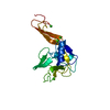
| ||||||||
|---|---|---|---|---|---|---|---|---|---|
| 1 |
| ||||||||
| Unit cell |
| ||||||||
| Atom site foot note | 1: THERE IS NO DENSITY FOR THE SIDE CHAIN OF RESIDUE GLU 8 BEYOND CB. 2: RESIDUE ASN 58 HAS A GENERALLY WEAK DENSITY BEYOND CB WITH ND2 TOTALLY OUT OF THE DENSITY. 3: ATOM NZ OF RESIDUE LYS 67 IS JUST OUT OF THE SIDE CHAIN DENSITY. 4: THE FOLLOWING ATOMS OF RESIDUE GLU 72 ARE OUT OF DENSITY: CD, OE1 AND OE2. 5: CIS PROLINE - PRO 81 6: THERE IS NO DENSITY FOR THE SIDE CHAIN OF RESIDUE ARG 84 BEYOND CB. 7: THERE IS WEAK DENSITY FOR THE SIDE CHAIN OF RESIDUE GLN 85 BEYOND CB. 8: THERE IS WEAK OR NO DENSITY FOR THE SIDE CHAIN OF RESIDUE GLU 98 BEYOND CB. 9: NO DENSITY FOUND FOR ATOM CD OF RESIDUE LYS 99. 10: RESIDUE ARG 108 HAS A BREAK IN THE DENSITY AT THE CG-CD BOND. 11: NO DENSITY FOUND FOR ATOM NZ OF RESIDUE LYS 112. 12: THE FOLLOWING ATOMS OF RESIDUE ASN 124 ARE OUT OF DENSITY: CG, OD1 AND OD2. 13: NO DENSITY FOUND FOR ATOM CG2 OF RESIDUE THR 125. 14: THERE IS WEAK DENSITY FOR THE SIDE CHAIN OF RESIDUE GLU 132 BEYOND CG. 15: THERE IS WEAK DENSITY FOR THE SIDE CHAIN OF RESIDUE LYS 143 BEYOND CG. 16: THERE IS WEAK DENSITY FOR THE C-TERMINAL CARBOXYLATE, RESIDUE VAL 157. |
-Components
| #1: Protein | Mass: 18605.820 Da / Num. of mol.: 1 Source method: isolated from a genetically manipulated source Source: (gene. exp.) Homo sapiens (human) / Cell line: CHO / References: UniProt: P16581 | ||||||
|---|---|---|---|---|---|---|---|
| #2: Chemical |   Mass: 40.078 Da / Num. of mol.: 3 / Source method: obtained synthetically / Formula: Ca Mass: 40.078 Da / Num. of mol.: 3 / Source method: obtained synthetically / Formula: Ca#3: Chemical | Chloride  Mass: 35.453 Da / Num. of mol.: 2 / Source method: obtained synthetically / Formula: Cl Mass: 35.453 Da / Num. of mol.: 2 / Source method: obtained synthetically / Formula: Cl#4: Water | ChemComp-HOH / | Water Mass: 18.015 Da / Num. of mol.: 112 / Source method: isolated from a natural source / Formula: H2O Mass: 18.015 Da / Num. of mol.: 112 / Source method: isolated from a natural source / Formula: H2ONonpolymer details | THREE WATER MOLECULES (HOH 260, HOH 266 AND HOH 318) HAVE TEMPERATURE FACTORS ABOVE 60 A**2 BUT ALL ...THREE WATER MOLECULES (HOH 260, HOH 266 AND HOH 318) HAVE TEMPERATUR | |
-Experimental details
-Experiment
| Experiment | Method: X-RAY DIFFRACTION |
|---|
- Sample preparation
| Crystal | Density Matthews: 2.64 Å3/Da / Density % sol: 53.32 % |
|---|---|
| Crystal grow | *PLUS Method: unknown |
-Data collection
| Radiation | Scattering type: x-ray |
|---|---|
| Radiation wavelength | Relative weight: 1 |
| Reflection | *PLUS Highest resolution: 2 Å / Rmerge(I) obs: 0.098 |
- Processing
| Software |
| ||||||||||||||||||||||||||||||||||||||||||||||||||||||||||||
|---|---|---|---|---|---|---|---|---|---|---|---|---|---|---|---|---|---|---|---|---|---|---|---|---|---|---|---|---|---|---|---|---|---|---|---|---|---|---|---|---|---|---|---|---|---|---|---|---|---|---|---|---|---|---|---|---|---|---|---|---|---|
| Refinement | Resolution: 2→10 Å / σ(F): 3
| ||||||||||||||||||||||||||||||||||||||||||||||||||||||||||||
| Refinement step | Cycle: LAST / Resolution: 2→10 Å
| ||||||||||||||||||||||||||||||||||||||||||||||||||||||||||||
| Refine LS restraints |
| ||||||||||||||||||||||||||||||||||||||||||||||||||||||||||||
| Software | *PLUS Name: X-PLOR / Classification: refinement | ||||||||||||||||||||||||||||||||||||||||||||||||||||||||||||
| Refinement | *PLUS Rfactor all: 0.173 / Rfactor obs: 0.164 / Rfactor Rfree: 0.249 | ||||||||||||||||||||||||||||||||||||||||||||||||||||||||||||
| Solvent computation | *PLUS | ||||||||||||||||||||||||||||||||||||||||||||||||||||||||||||
| Displacement parameters | *PLUS | ||||||||||||||||||||||||||||||||||||||||||||||||||||||||||||
| Refine LS restraints | *PLUS Type: x_angle_d / Dev ideal: 2.6 |
