 Movie
Movie Controller

[English] 日本語
 Yorodumi
- PDB-3cdm: Structural adaptation and conservation in quadruplex-drug recognition -
+ Open data

- Basic information
| Entry | Database: PDB / ID: 3cdm | ||||||||||||||||||
|---|---|---|---|---|---|---|---|---|---|---|---|---|---|---|---|---|---|---|---|
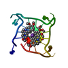
| Title | Structural adaptation and conservation in quadruplex-drug recognition | ||||||||||||||||||
 Components Components | DNA (5'-D(* Keywords Keywords DNA / QUADRUPLEX / PROPELLER / INTRAMOLECULAR / HUMAN TELOMERE PARALLEL STRANDED / LIGAND / COMPLEX DNA / QUADRUPLEX / PROPELLER / INTRAMOLECULAR / HUMAN TELOMERE PARALLEL STRANDED / LIGAND / COMPLEXFunction / homology | : / Chem-NII / DNA / DNA (> 10) Function and homology information Function and homology informationMethod | X-RAY DIFFRACTION / SYNCHROTRON / MOLECULAR REPLACEMENT / Resolution: 2.1 Å  Authors AuthorsParkinson, G.N. / Neidle, S. |  CitationJournal: J.Mol.Biol. / Year: 2008 CitationJournal: J.Mol.Biol. / Year: 2008Title: Topology conservation and loop flexibility in quadruplex-drug recognition: crystal structures of inter- and intramolecular telomeric DNA quadruplex-drug complexes Authors: Parkinson, G.N. / Cuenca, F. / Neidle, S. History |
|
- Structure visualization
| Structure viewer | Molecule: MolmilJmol/JSmol |
|---|
- Downloads & links
-Download
| PDBx/mmCIF format | 3cdm.cif.gz | 48.9 KB | Display | PDBx/mmCIF format |
|---|---|---|---|---|
| PDB format | pdb3cdm.ent.gz | 35.1 KB | Display | PDB format |
| PDBx/mmJSON format | 3cdm.json.gz | Tree view | PDBx/mmJSON format | |
| Others |  Other downloads Other downloads |
-Validation report
| Arichive directory | https://data.pdbj.org/pub/pdb/validation_reports/cd/3cdmftp://data.pdbj.org/pub/pdb/validation_reports/cd/3cdm | HTTPS FTP |
|---|
-Related structure data
| Related structure data | 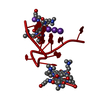 3ccoC  1kf1S C: citing same article ( S: Starting model for refinement |
|---|---|
| Similar structure data |


-Links
 PDBj
PDBj


- Assembly
| Deposited unit | 
| ||||||||
|---|---|---|---|---|---|---|---|---|---|
| 1 | 
| ||||||||
| 2 | 
| ||||||||
| Unit cell |
|
-Components
| #1: DNA chain | Mass: 7287.690 Da / Num. of mol.: 2 / Source method: obtained synthetically / Details: This sequence occurs naturally in humans #2: Chemical | ChemComp-NII /   Mass: 582.691 Da / Num. of mol.: 6 / Source method: obtained synthetically / Formula: C30H42N6O6 Mass: 582.691 Da / Num. of mol.: 6 / Source method: obtained synthetically / Formula: C30H42N6O6#3: Chemical | ChemComp-K /   Mass: 39.098 Da / Num. of mol.: 4 / Source method: obtained synthetically / Formula: K Mass: 39.098 Da / Num. of mol.: 4 / Source method: obtained synthetically / Formula: K#4: Water | ChemComp-HOH / | Water Mass: 18.015 Da / Num. of mol.: 158 / Source method: isolated from a natural source / Formula: H2O Mass: 18.015 Da / Num. of mol.: 158 / Source method: isolated from a natural source / Formula: H2O |
|---|
-Experimental details
-Experiment
| Experiment | Method: X-RAY DIFFRACTION / Number of used crystals: 1 |
|---|
- Sample preparation
| Crystal | Density Matthews: 3.24 Å3/Da / Density % sol: 62 % | ||||||||||||||||||||||||||||||||||||||||||||||||||||
|---|---|---|---|---|---|---|---|---|---|---|---|---|---|---|---|---|---|---|---|---|---|---|---|---|---|---|---|---|---|---|---|---|---|---|---|---|---|---|---|---|---|---|---|---|---|---|---|---|---|---|---|---|---|
| Crystal grow | Temperature: 283 K / Method: vapor diffusion, hanging drop / pH: 6.5 Details: 333mM ammonium sulfate, 10mM magnesium chloride, 50mM sodium chloride, 50mM potassium chloride, 50mM potassium cacodylate pH 6.5, VAPOR DIFFUSION, HANGING DROP, temperature 283K | ||||||||||||||||||||||||||||||||||||||||||||||||||||
| Components of the solutions |
|
-Data collection
| Diffraction | Mean temperature: 100 K |
|---|---|
| Diffraction source | Source: SYNCHROTRON / Site: ESRF  / Beamline: ID14-1 / Wavelength: 0.9785 Å / Beamline: ID14-1 / Wavelength: 0.9785 Å |
| Detector | Type: ADSC QUANTUM 210 / Detector: CCD / Date: Oct 7, 2007 / Details: Monochromator |
| Radiation | Monochromator: Si 111 CHANNEL / Protocol: SINGLE WAVELENGTH / Monochromatic (M) / Laue (L): M / Scattering type: x-ray |
| Radiation wavelength | Wavelength: 0.9785 Å / Relative weight: 1 |
| Reflection | Resolution: 2.1→28.69 Å / Num. all: 10055 / Num. obs: 10055 / % possible obs: 91 % / Observed criterion σ(I): 0 / Redundancy: 2.86 % / Biso Wilson estimate: 46 Å2 / Rmerge(I) obs: 0.036 / Net I/σ(I): 17.9 |
| Reflection shell | Resolution: 2.1→2.18 Å / Redundancy: 2.94 % / Rmerge(I) obs: 0.116 / Mean I/σ(I) obs: 6.4 / Num. unique all: 1041 / % possible all: 95.9 |
- Processing
| Software |
| |||||||||||||||||||||||||||||||||||||||||||||||||||||||||||||||||
|---|---|---|---|---|---|---|---|---|---|---|---|---|---|---|---|---|---|---|---|---|---|---|---|---|---|---|---|---|---|---|---|---|---|---|---|---|---|---|---|---|---|---|---|---|---|---|---|---|---|---|---|---|---|---|---|---|---|---|---|---|---|---|---|---|---|---|
| Refinement | Method to determine structure: MOLECULAR REPLACEMENT Starting model: PDB ENTRY 1KF1 Resolution: 2.1→10 Å / Cor.coef. Fo:Fc: 0.936 / Cor.coef. Fo:Fc free: 0.886 / SU B: 5.422 / SU ML: 0.152 / Isotropic thermal model: Overall / Cross valid method: THROUGHOUT / σ(F): 0 / ESU R: 0.295 / ESU R Free: 0.246 / Stereochemistry target values: MAXIMUM LIKELIHOOD
| |||||||||||||||||||||||||||||||||||||||||||||||||||||||||||||||||
| Displacement parameters | Biso mean: 26.776 Å2
| |||||||||||||||||||||||||||||||||||||||||||||||||||||||||||||||||
| Refine analyze | Luzzati coordinate error obs: 0.267 Å | |||||||||||||||||||||||||||||||||||||||||||||||||||||||||||||||||
| Refinement step | Cycle: LAST / Resolution: 2.1→10 Å
| |||||||||||||||||||||||||||||||||||||||||||||||||||||||||||||||||
| Refine LS restraints |
| |||||||||||||||||||||||||||||||||||||||||||||||||||||||||||||||||
| LS refinement shell | Resolution: 2.1→2.152 Å / Total num. of bins used: 20
|
