 Movie
Movie Controller


+ Open data

- Basic information
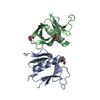
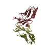
| Entry | Database: PDB / ID: 3cbx | ||||||
|---|---|---|---|---|---|---|---|
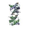
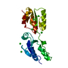
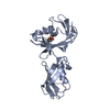
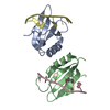
| Title | The Dvl2 PDZ Domain in Complex with the C1 Inhibitory Peptide | ||||||
 Components Components | Dishevelled-2 | ||||||
 Keywords Keywords | PROTEIN BINDING / PDZ DOMAIN / PHAGE DERIVED HIGH AFFINITY LIGAND / Developmental protein / Phosphoprotein / Wnt signaling pathway / SIGNALING PROTEIN | ||||||
| Function / homology |  Function and homology information Function and homology informationNegative regulation of TCF-dependent signaling by DVL-interacting proteins / convergent extension involved in neural plate elongation / planar cell polarity pathway involved in neural tube closure / cochlea morphogenesis / segment specification / WNT5A-dependent internalization of FZD4 / non-canonical Wnt signaling pathway / positive regulation of neuron projection arborization / WNT5:FZD7-mediated leishmania damping / clathrin-coated endocytic vesicle ...Negative regulation of TCF-dependent signaling by DVL-interacting proteins / convergent extension involved in neural plate elongation / planar cell polarity pathway involved in neural tube closure / cochlea morphogenesis / segment specification / WNT5A-dependent internalization of FZD4 / non-canonical Wnt signaling pathway / positive regulation of neuron projection arborization / WNT5:FZD7-mediated leishmania damping / clathrin-coated endocytic vesicle / frizzled binding / PCP/CE pathway / Signaling by Hippo / WNT mediated activation of DVL / aggresome / Disassembly of the destruction complex and recruitment of AXIN to the membrane / Wnt signaling pathway, planar cell polarity pathway / heart looping / outflow tract morphogenesis / lateral plasma membrane / canonical Wnt signaling pathway / positive regulation of JUN kinase activity / Asymmetric localization of PCP proteins / TCF dependent signaling in response to WNT / neural tube closure / RHO GTPases Activate Formins / Degradation of DVL / regulation of actin cytoskeleton organization / positive regulation of JNK cascade / protein localization / small GTPase binding / positive regulation of DNA-binding transcription factor activity / : / Cargo recognition for clathrin-mediated endocytosis / apical part of cell / protein-macromolecule adaptor activity / Clathrin-mediated endocytosis / heart development / regulation of cell population proliferation / cytoplasmic vesicle / nuclear body / intracellular signal transduction / positive regulation of protein phosphorylation / protein domain specific binding / regulation of DNA-templated transcription / protein kinase binding / positive regulation of transcription by RNA polymerase II / nucleoplasm / identical protein binding / nucleus / cytosol / cytoplasmSimilarity search - Function | ||||||
| Biological species |  Homo sapiens (human) Homo sapiens (human) | ||||||
| Method | X-RAY DIFFRACTION / SYNCHROTRON / MOLECULAR REPLACEMENT / molecular replacement / Resolution: 1.7 Å | ||||||
 Authors Authors | Appleton, B.A. / Wiesmann, C. | ||||||
 Citation Citation | Journal: Nat.Chem.Biol. / Year: 2009 Title: Inhibition of Wnt signaling by Dishevelled PDZ peptides Authors: Zhang, Y. / Appleton, B.A. / Wiesmann, C. / Lau, T. / Costa, M. / Hannoush, R.N. / Sidhu, S.S. | ||||||
| History |
|
- Structure visualization
| Structure viewer | Molecule: MolmilJmol/JSmol |
|---|
- Downloads & links
-Download
| PDBx/mmCIF format | 3cbx.cif.gz | 56.9 KB | Display | PDBx/mmCIF format |
|---|---|---|---|---|
| PDB format | pdb3cbx.ent.gz | 40.2 KB | Display | PDB format |
| PDBx/mmJSON format | 3cbx.json.gz | Tree view | PDBx/mmJSON format | |
| Others |  Other downloads Other downloads |
-Validation report
| Arichive directory | https://data.pdbj.org/pub/pdb/validation_reports/cb/3cbxftp://data.pdbj.org/pub/pdb/validation_reports/cb/3cbx | HTTPS FTP |
|---|
-Related structure data
| Related structure data |  3cbyC 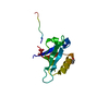 3cbzC 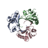 3cc0C 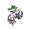 1l6oS C: citing same article ( S: Starting model for refinement |
|---|---|
| Similar structure data |

-Links
 PDBj
PDBj


- Assembly
| Deposited unit | 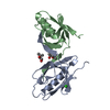
| ||||||||
|---|---|---|---|---|---|---|---|---|---|
| 1 |
| ||||||||
| Unit cell |
|
-Components
| #1: Protein | / DSH homolog 2 / Segment polarity protein dishevelled homolog DVL-2 Mass: 11425.021 Da / Num. of mol.: 2 / Fragment: PDZ domain (UNP residues 264-354) / Mutation: C341S Source method: isolated from a genetically manipulated source Source: (gene. exp.) Homo sapiens (human) / Gene: DVL2 / Production host:  Escherichia coli (E. coli) / References: UniProt: O14641 Escherichia coli (E. coli) / References: UniProt: O14641#2: Chemical | 2-Methyl-2,4-pentanediol  Mass: 118.174 Da / Num. of mol.: 2 / Source method: obtained synthetically / Formula: C6H14O2 / Comment: precipitant*YM Mass: 118.174 Da / Num. of mol.: 2 / Source method: obtained synthetically / Formula: C6H14O2 / Comment: precipitant*YM#3: Chemical | ChemComp-CL / | Chloride  Mass: 35.453 Da / Num. of mol.: 1 / Source method: obtained synthetically / Formula: Cl Mass: 35.453 Da / Num. of mol.: 1 / Source method: obtained synthetically / Formula: Cl#4: Water | ChemComp-HOH / | Water Mass: 18.015 Da / Num. of mol.: 121 / Source method: isolated from a natural source / Formula: H2O Mass: 18.015 Da / Num. of mol.: 121 / Source method: isolated from a natural source / Formula: H2OSequence details | THE PEPTIDE LIGAND WAS FUSED TO THE C TERMINUS OF THE LINKER | |
|---|
-Experimental details
-Experiment
| Experiment | Method: X-RAY DIFFRACTION / Number of used crystals: 1 |
|---|
- Sample preparation
| Crystal | Density Matthews: 2.25 Å3/Da / Density % sol: 45.42 % |
|---|---|
| Crystal grow | Temperature: 292 K / pH: 4.8 Details: 60% 2-Methyl-2,4-pentanediol, 0.1 M sodium acetate, pH 4.8, VAPOR DIFFUSION, SITTING DROP, temperature 292K |
-Data collection
| Diffraction | Mean temperature: 100 K |
|---|---|
| Diffraction source | Source: SYNCHROTRON / Site: ALS  / Beamline: 5.0.2 / Wavelength: 1 / Beamline: 5.0.2 / Wavelength: 1 |
| Detector | Type: ADSC QUANTUM 315 / Detector: CCD / Date: Feb 15, 2006 |
| Radiation | Protocol: SINGLE WAVELENGTH / Monochromatic (M) / Laue (L): M / Scattering type: x-ray |
| Radiation wavelength | Wavelength: 1 Å / Relative weight: 1 |
| Reflection | Resolution: 1.7→50 Å / Num. obs: 23342 / % possible obs: 99.6 % / Redundancy: 4.6 % / Rmerge(I) obs: 0.069 / Net I/σ(I): 16.9 |
| Reflection shell | Resolution: 1.7→1.76 Å / Redundancy: 4.5 % / Rmerge(I) obs: 0.463 / Mean I/σ(I) obs: 3.2 / % possible all: 99.9 |
-Phasing
| Phasing | Method: molecular replacement |
|---|
- Processing
| Software |
| ||||||||||||||||||||||||||||||||||||||||||||||||||||||||||||||||||||||||||||||||||||||||||||||||||||||||||||||||||||||||||||||||||||||||||||||||||||||||||||||||||||||||||
|---|---|---|---|---|---|---|---|---|---|---|---|---|---|---|---|---|---|---|---|---|---|---|---|---|---|---|---|---|---|---|---|---|---|---|---|---|---|---|---|---|---|---|---|---|---|---|---|---|---|---|---|---|---|---|---|---|---|---|---|---|---|---|---|---|---|---|---|---|---|---|---|---|---|---|---|---|---|---|---|---|---|---|---|---|---|---|---|---|---|---|---|---|---|---|---|---|---|---|---|---|---|---|---|---|---|---|---|---|---|---|---|---|---|---|---|---|---|---|---|---|---|---|---|---|---|---|---|---|---|---|---|---|---|---|---|---|---|---|---|---|---|---|---|---|---|---|---|---|---|---|---|---|---|---|---|---|---|---|---|---|---|---|---|---|---|---|---|---|---|---|---|
| Refinement | Method to determine structure: MOLECULAR REPLACEMENT Starting model: 1L6O Resolution: 1.7→20 Å / Cor.coef. Fo:Fc: 0.962 / Cor.coef. Fo:Fc free: 0.942 / SU B: 4.417 / SU ML: 0.076 / TLS residual ADP flag: LIKELY RESIDUAL / Cross valid method: THROUGHOUT / σ(F): 0 / ESU R: 0.108 / ESU R Free: 0.11 / Stereochemistry target values: MAXIMUM LIKELIHOOD / Details: HYDROGENS HAVE BEEN ADDED IN THE RIDING POSITIONS
| ||||||||||||||||||||||||||||||||||||||||||||||||||||||||||||||||||||||||||||||||||||||||||||||||||||||||||||||||||||||||||||||||||||||||||||||||||||||||||||||||||||||||||
| Solvent computation | Ion probe radii: 0.8 Å / Shrinkage radii: 0.8 Å / VDW probe radii: 1.4 Å / Solvent model: BABINET MODEL WITH MASK | ||||||||||||||||||||||||||||||||||||||||||||||||||||||||||||||||||||||||||||||||||||||||||||||||||||||||||||||||||||||||||||||||||||||||||||||||||||||||||||||||||||||||||
| Displacement parameters | Biso mean: 27.92 Å2
| ||||||||||||||||||||||||||||||||||||||||||||||||||||||||||||||||||||||||||||||||||||||||||||||||||||||||||||||||||||||||||||||||||||||||||||||||||||||||||||||||||||||||||
| Refinement step | Cycle: LAST / Resolution: 1.7→20 Å
| ||||||||||||||||||||||||||||||||||||||||||||||||||||||||||||||||||||||||||||||||||||||||||||||||||||||||||||||||||||||||||||||||||||||||||||||||||||||||||||||||||||||||||
| Refine LS restraints |
| ||||||||||||||||||||||||||||||||||||||||||||||||||||||||||||||||||||||||||||||||||||||||||||||||||||||||||||||||||||||||||||||||||||||||||||||||||||||||||||||||||||||||||
| LS refinement shell | Resolution: 1.7→1.74 Å / Total num. of bins used: 25
| ||||||||||||||||||||||||||||||||||||||||||||||||||||||||||||||||||||||||||||||||||||||||||||||||||||||||||||||||||||||||||||||||||||||||||||||||||||||||||||||||||||||||||
| Refinement TLS params. | Method: refined / Refine-ID: X-RAY DIFFRACTION
| ||||||||||||||||||||||||||||||||||||||||||||||||||||||||||||||||||||||||||||||||||||||||||||||||||||||||||||||||||||||||||||||||||||||||||||||||||||||||||||||||||||||||||
| Refinement TLS group |
|
