- PDB-3c6h: Crystal Structure of the RB49 gp17 nuclease domain -
+
Open data
ID or keywords:
Loading...
-
Basic information
Entry
Database: PDB / ID: 3c6h
Title
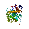
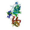
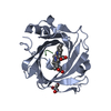
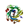
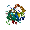
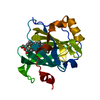
Crystal Structure of the RB49 gp17 nuclease domain
Components
Terminase large subunit
Keywords
VIRAL PROTEIN / terminase nuclease
Function / homology
Function and homology information
viral terminase, large subunit / viral DNA genome packaging / Hydrolases; Acting on ester bonds; Endodeoxyribonucleases producing 5'-phosphomonoesters / chromosome organization / Hydrolases; Acting on acid anhydrides; Acting on acid anhydrides to facilitate cellular and subcellular movement / endonuclease activity / ATP hydrolysis activity / ATP binding / metal ion binding Similarity search - Function
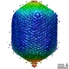
Journal: Cell / Year: 2008 Title: The structure of the phage T4 DNA packaging motor suggests a mechanism dependent on electrostatic forces. Authors: Siyang Sun / Kiran Kondabagil / Bonnie Draper / Tanfis I Alam / Valorie D Bowman / Zhihong Zhang / Shylaja Hegde / Andrei Fokine / Michael G Rossmann / Venigalla B Rao / Abstract: Viral genomes are packaged into "procapsids" by powerful molecular motors. We report the crystal structure of the DNA packaging motor protein, gene product 17 (gp17), in bacteriophage T4. The ...Viral genomes are packaged into "procapsids" by powerful molecular motors. We report the crystal structure of the DNA packaging motor protein, gene product 17 (gp17), in bacteriophage T4. The structure consists of an N-terminal ATPase domain, which provides energy for compacting DNA, and a C-terminal nuclease domain, which terminates packaging. We show that another function of the C-terminal domain is to translocate the genome into the procapsid. The two domains are in close contact in the crystal structure, representing a "tensed state." A cryo-electron microscopy reconstruction of the T4 procapsid complexed with gp17 shows that the packaging motor is a pentamer and that the domains within each monomer are spatially separated, representing a "relaxed state." These structures suggest a mechanism, supported by mutational and other data, in which electrostatic forces drive the DNA packaging by alternating between tensed and relaxed states. Similar mechanisms may occur in other molecular motors.
In the structure databanks used in Yorodumi, some data are registered as the other names, "COVID-19 virus" and "2019-nCoV". Here are the details of the virus and the list of structure data.
Jan 31, 2019. EMDB accession codes are about to change! (news from PDBe EMDB page)
EMDB accession codes are about to change! (news from PDBe EMDB page)
The allocation of 4 digits for EMDB accession codes will soon come to an end. Whilst these codes will remain in use, new EMDB accession codes will include an additional digit and will expand incrementally as the available range of codes is exhausted. The current 4-digit format prefixed with “EMD-” (i.e. EMD-XXXX) will advance to a 5-digit format (i.e. EMD-XXXXX), and so on. It is currently estimated that the 4-digit codes will be depleted around Spring 2019, at which point the 5-digit format will come into force.
The EM Navigator/Yorodumi systems omit the EMD- prefix.
Related info.:Q: What is EMD? / ID/Accession-code notation in Yorodumi/EM Navigator
Yorodumi is a browser for structure data from EMDB, PDB, SASBDB, etc.
This page is also the successor to EM Navigator detail page, and also detail information page/front-end page for Omokage search.
The word "yorodu" (or yorozu) is an old Japanese word meaning "ten thousand". "mi" (miru) is to see.
Related info.:EMDB / PDB / SASBDB / Comparison of 3 databanks / Yorodumi Search / Aug 31, 2016. New EM Navigator & Yorodumi / Yorodumi Papers / Jmol/JSmol / Function and homology information / Changes in new EM Navigator and Yorodumi
 Movie
Movie Controller


 Open data
 Basic information
 Components
 Keywords
 VIRAL PROTEIN / terminase nuclease
 Function and homology information
 Enterobacteria phage RB49 (virus)
 Authors
 Citation
 Structure visualization
 Downloads & links
 Other downloads


 PDBj
PDBj Assembly


 Mass: 24.305 Da / Num. of mol.: 2 / Source method: obtained synthetically / Formula: Mg
 Mass: 18.015 Da / Num. of mol.: 96 / Source method: isolated from a natural source / Formula: H2O
 Sample preparation
 Processing