[English] 日本語
Yorodumi
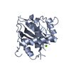
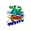
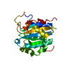
- PDB-3c6a: Crystal Structure of the RB49 gp17 nuclease domain -

+
Open data


ID or keywords:

Loading...

-
Basic information

Entry
Database: PDB / ID: 3c6a
TitleCrystal Structure of the RB49 gp17 nuclease domain
ComponentsTerminase large subunit
KeywordsVIRAL PROTEIN / terminase nuclease
Function / homology
Function and homology information


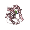
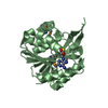
viral terminase, large subunit / viral DNA genome packaging / Hydrolases; Acting on ester bonds; Endodeoxyribonucleases producing 5'-phosphomonoesters / chromosome organization / Hydrolases; Acting on acid anhydrides; Acting on acid anhydrides to facilitate cellular and subcellular movement / endonuclease activity / ATP hydrolysis activity / ATP binding / metal ion binding
Similarity search - Function
Nucleotidyltransferase; domain 5 - #240 / Terminase, large subunit, gp17-like / Terminase, large subunit gp17-like, C-terminal / Terminase RNaseH-like domain / Terminase large subunit, T4likevirus-type, N-terminal / Nucleotidyltransferase; domain 5 / P-loop containing nucleoside triphosphate hydrolase / 2-Layer Sandwich / Alpha Beta
Similarity search - Domain/homology
Terminase, large subunit
Similarity search - Component
Biological speciesEnterobacteria phage RB49 (virus)
MethodX-RAY DIFFRACTION / SYNCHROTRON / AB INITIO PHASING / SIRAS / Resolution: 1.16 Å
AuthorsSun, S. / Rossmann, M.G.
Citation
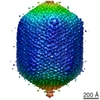
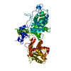
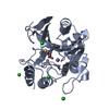
Journal: Cell / Year: 2008
Title: The structure of the phage T4 DNA packaging motor suggests a mechanism dependent on electrostatic forces.
Authors: Siyang Sun / Kiran Kondabagil / Bonnie Draper / Tanfis I Alam / Valorie D Bowman / Zhihong Zhang / Shylaja Hegde / Andrei Fokine / Michael G Rossmann / Venigalla B Rao /
Abstract: Viral genomes are packaged into "procapsids" by powerful molecular motors. We report the crystal structure of the DNA packaging motor protein, gene product 17 (gp17), in bacteriophage T4. The ...Viral genomes are packaged into "procapsids" by powerful molecular motors. We report the crystal structure of the DNA packaging motor protein, gene product 17 (gp17), in bacteriophage T4. The structure consists of an N-terminal ATPase domain, which provides energy for compacting DNA, and a C-terminal nuclease domain, which terminates packaging. We show that another function of the C-terminal domain is to translocate the genome into the procapsid. The two domains are in close contact in the crystal structure, representing a "tensed state." A cryo-electron microscopy reconstruction of the T4 procapsid complexed with gp17 shows that the packaging motor is a pentamer and that the domains within each monomer are spatially separated, representing a "relaxed state." These structures suggest a mechanism, supported by mutational and other data, in which electrostatic forces drive the DNA packaging by alternating between tensed and relaxed states. Similar mechanisms may occur in other molecular motors.
History
DepositionFeb 4, 2008Deposition site: RCSB / Processing site: RCSB
Revision 1.0Jan 13, 2009Provider: repository / Type: Initial release
Revision 1.1Jul 13, 2011Group: Version format compliance
Revision 1.2Oct 25, 2017Group: Refinement description / Category: software
Item: _software.classification / _software.contact_author ..._software.classification / _software.contact_author / _software.contact_author_email / _software.date / _software.language / _software.location / _software.name / _software.type / _software.version
Revision 1.3Feb 21, 2024Group: Data collection / Database references / Derived calculations
Category: chem_comp_atom / chem_comp_bond ...chem_comp_atom / chem_comp_bond / database_2 / struct_ref_seq_dif / struct_site
Item: _database_2.pdbx_DOI / _database_2.pdbx_database_accession ..._database_2.pdbx_DOI / _database_2.pdbx_database_accession / _struct_ref_seq_dif.details / _struct_site.pdbx_auth_asym_id / _struct_site.pdbx_auth_comp_id / _struct_site.pdbx_auth_seq_id

-
Structure visualization

Structure viewerMolecule:
MolmilJmol/JSmol

Downloads & links

-
Assembly

Deposited unit
A: Terminase large subunit
hetero molecules


Theoretical massNumber of molelcules
Total (without water)26,3662
Polymers26,3421
Non-polymers241
Water6,990388
1


  • Idetical with deposited unit
  • defined by author&software
TypeNameSymmetry operationNumber
identity operation1_555x,y,z1
MethodPISA
Unit cell
Length a, b, c (Å)52.568, 125.163, 37.218
Angle α, β, γ (deg.)90.000, 90.000, 90.000
Int Tables number18
Space group name H-MP21212

-
Components

#1: Protein Terminase large subunit


Mass: 26341.957 Da / Num. of mol.: 1 / Fragment: nuclease domain (UNP residues 359-564)
Source method: isolated from a genetically manipulated source
Source: (gene. exp.) Enterobacteria phage RB49 (virus) / Gene: 17 / Plasmid: pET28 / Production host: Escherichia coli (E. coli) / Strain (production host): BL21(DE3) pLysS / References: UniProt: Q9T1C3
#2: Chemical ChemComp-MG / MAGNESIUM ION


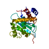
Mass: 24.305 Da / Num. of mol.: 1 / Source method: obtained synthetically / Formula: Mg
#3: Water ChemComp-HOH / water / Water


Mass: 18.015 Da / Num. of mol.: 388 / Source method: isolated from a natural source / Formula: H2O

-
Experimental details

-
Experiment

ExperimentMethod: X-RAY DIFFRACTION / Number of used crystals: 1

-
Sample preparation

CrystalDensity Matthews: 2.32 Å3/Da / Density % sol: 47.07 %
Crystal growTemperature: 293 K / Method: vapor diffusion, hanging drop / pH: 7
Details: 15-20% PEG3350, 0.2M Na/K tartrate, pH 7.0, VAPOR DIFFUSION, HANGING DROP, temperature 293K

-
Data collection

DiffractionMean temperature: 100 K
Diffraction sourceSource: SYNCHROTRON / Site: APS / Beamline: 23-ID-B / Wavelength: 1.033 Å
DetectorType: MARMOSAIC 300 mm CCD / Detector: CCD / Date: Oct 1, 2007 / Details: mirrors
RadiationMonochromator: GRAPHITE / Protocol: SINGLE WAVELENGTH / Monochromatic (M) / Laue (L): M / Scattering type: x-ray
Radiation wavelengthWavelength: 1.033 Å / Relative weight: 1
ReflectionRedundancy: 4.6 % / Av σ(I) over netI: 23.3 / Number: 149604 / Rmerge(I) obs: 0.033 / Χ2: 1.57 / D res high: 1.6 Å / D res low: 50 Å / Num. obs: 32564 / % possible obs: 97.6
Diffraction reflection shell
Highest resolution (Å)Lowest resolution (Å)% possible obs (%)IDRmerge(I) obsChi squaredRedundancy
3.455098.810.0251.1984.5
2.743.4599.810.0231.1684.7
2.392.7499.610.0291.3014.7
2.172.3998.810.0361.54.7
2.022.1798.710.0451.5934.7
1.92.0297.810.0621.7564.7
1.81.997.510.0881.8234.7
1.721.896.910.1241.8494.6
1.661.7296.510.1661.844.6
1.61.6690.910.2181.8363.9
ReflectionResolution: 1.16→50 Å / Num. all: 86090 / Num. obs: 85573 / % possible obs: 99.4 % / Observed criterion σ(F): 0 / Observed criterion σ(I): 0 / Redundancy: 5.8 % / Biso Wilson estimate: 10.8 Å2 / Rmerge(I) obs: 0.041 / Χ2: 1.18 / Net I/σ(I): 12.7
Reflection shellResolution: 1.16→1.2 Å / Redundancy: 5.1 % / Rmerge(I) obs: 0.384 / Mean I/σ(I) obs: 3.9 / Num. unique all: 8226 / Χ2: 0.939 / % possible all: 96.6

-
Phasing

PhasingMethod: SIRAS
Phasing MAD set
IDR cullis acentricR cullis centricHighest resolution (Å)Lowest resolution (Å)Reflection acentricReflection centric
ISO_1001.620.3286793434
ISO_20.6420.5991.620.34021469
ANO_1001.620.300
ANO_20.73301.620.339880
Phasing MAD set shell
IDResolution (Å)R cullis acentricR cullis centricReflection acentricReflection centric
ISO_16.77-20.300299180
ISO_14.92-6.7700553184
ISO_14.06-4.9200732179
ISO_13.53-4.0600877183
ISO_13.17-3.53001024180
ISO_12.9-3.17001120183
ISO_12.69-2.9001228182
ISO_12.52-2.69001334170
ISO_12.38-2.52001413174
ISO_12.26-2.38001498162
ISO_12.15-2.26001574165
ISO_12.06-2.15001635155
ISO_11.98-2.06001725163
ISO_11.91-1.98001800177
ISO_11.85-1.91001853170
ISO_11.79-1.85001909168
ISO_11.73-1.79001982185
ISO_11.69-1.73002015175
ISO_11.64-1.69002086167
ISO_11.6-1.64002022132
ANO_16.77-20.30000
ANO_14.92-6.770000
ANO_14.06-4.920000
ANO_13.53-4.060000
ANO_13.17-3.530000
ANO_12.9-3.170000
ANO_12.69-2.90000
ANO_12.52-2.690000
ANO_12.38-2.520000
ANO_12.26-2.380000
ANO_12.15-2.260000
ANO_12.06-2.150000
ANO_11.98-2.060000
ANO_11.91-1.980000
ANO_11.85-1.910000
ANO_11.79-1.850000
ANO_11.73-1.790000
ANO_11.69-1.730000
ANO_11.64-1.690000
ANO_11.6-1.640000
ISO_26.77-20.30.5350.48427683
ISO_24.92-6.770.5910.67552785
ISO_24.06-4.920.6280.71770977
ISO_23.53-4.060.7040.61785290
ISO_23.17-3.530.720.723100281
ISO_22.9-3.170.7260.78665553
ISO_22.69-2.90000
ISO_22.52-2.690000
ISO_22.38-2.520000
ISO_22.26-2.380000
ISO_22.15-2.260000
ISO_22.06-2.150000
ISO_21.98-2.060000
ISO_21.91-1.980000
ISO_21.85-1.910000
ISO_21.79-1.850000
ISO_21.73-1.790000
ISO_21.69-1.730000
ISO_21.64-1.690000
ISO_21.6-1.640000
ANO_26.77-20.30.63502720
ANO_24.92-6.770.70105210
ANO_24.06-4.920.72107000
ANO_23.53-4.060.73308450
ANO_23.17-3.530.81409990
ANO_22.9-3.170.84106510
ANO_22.69-2.90000
ANO_22.52-2.690000
ANO_22.38-2.520000
ANO_22.26-2.380000
ANO_22.15-2.260000
ANO_22.06-2.150000
ANO_21.98-2.060000
ANO_21.91-1.980000
ANO_21.85-1.910000
ANO_21.79-1.850000
ANO_21.73-1.790000
ANO_21.69-1.730000
ANO_21.64-1.690000
ANO_21.6-1.640000

-
Processing

Software
NameVersionClassificationNB
DENZOdata reduction
SCALEPACKdata scaling
SHARPphasing
SOLOMONphasing
SHELXrefinement
PDB_EXTRACT3.004data extraction
SHELXL-97refinement
RefinementMethod to determine structure: AB INITIO PHASING / Resolution: 1.16→10 Å / Num. parameters: 18219 / Num. restraintsaints: 21916 / Cross valid method: FREE R / σ(F): 0 / Stereochemistry target values: Engh & Huber
RfactorNum. reflection% reflectionSelection details
Rfree0.171 4298 -RANDOM
Rwork0.134 ---
obs0.134 81068 94.5 %-
all-85786 --
Solvent computationSolvent model: MOEWS & KRETSINGER, J.MOL.BIOL.91(1973)201-228
Displacement parametersBiso mean: 21.939 Å2
Refinement stepCycle: LAST / Resolution: 1.16→10 Å
ProteinNucleic acidLigandSolventTotal
Num. atoms1635 0 1 388 2024
Refine LS restraints
Refine-IDTypeDev ideal
X-RAY DIFFRACTIONs_bond_d0.017
X-RAY DIFFRACTIONs_angle_d0.031
X-RAY DIFFRACTIONs_similar_dist0
X-RAY DIFFRACTIONs_from_restr_planes0.027
X-RAY DIFFRACTIONs_zero_chiral_vol0.071
X-RAY DIFFRACTIONs_non_zero_chiral_vol0.095
X-RAY DIFFRACTIONs_anti_bump_dis_restr0.024
X-RAY DIFFRACTIONs_rigid_bond_adp_cmpnt0.006
X-RAY DIFFRACTIONs_similar_adp_cmpnt0.057
X-RAY DIFFRACTIONs_approx_iso_adps0.11

+
About Yorodumi

-
News

-
Feb 9, 2022. New format data for meta-information of EMDB entries

New format data for meta-information of EMDB entries

  • Version 3 of the EMDB header file is now the official format.
  • The previous official version 1.9 will be removed from the archive.

Related info.:EMDB header

External links:wwPDB to switch to version 3 of the EMDB data model

-
Aug 12, 2020. Covid-19 info

Covid-19 info

URL: https://pdbj.org/emnavi/covid19.php

New page: Covid-19 featured information page in EM Navigator.

Related info.:Covid-19 info / Mar 5, 2020. Novel coronavirus structure data

+
Mar 5, 2020. Novel coronavirus structure data

Novel coronavirus structure data

Related info.:Yorodumi Speices / Aug 12, 2020. Covid-19 info

External links:COVID-19 featured content - PDBj / Molecule of the Month (242):Coronavirus Proteases

+
Jan 31, 2019. EMDB accession codes are about to change! (news from PDBe EMDB page)

EMDB accession codes are about to change! (news from PDBe EMDB page)

  • The allocation of 4 digits for EMDB accession codes will soon come to an end. Whilst these codes will remain in use, new EMDB accession codes will include an additional digit and will expand incrementally as the available range of codes is exhausted. The current 4-digit format prefixed with “EMD-” (i.e. EMD-XXXX) will advance to a 5-digit format (i.e. EMD-XXXXX), and so on. It is currently estimated that the 4-digit codes will be depleted around Spring 2019, at which point the 5-digit format will come into force.
  • The EM Navigator/Yorodumi systems omit the EMD- prefix.

Related info.:Q: What is EMD? / ID/Accession-code notation in Yorodumi/EM Navigator

External links:EMDB Accession Codes are Changing Soon! / Contact to PDBj

+
Jul 12, 2017. Major update of PDB

Major update of PDB

  • wwPDB released updated PDB data conforming to the new PDBx/mmCIF dictionary.
  • This is a major update changing the version number from 4 to 5, and with Remediation, in which all the entries are updated.
  • In this update, many items about electron microscopy experimental information are reorganized (e.g. em_software).
  • Now, EM Navigator and Yorodumi are based on the updated data.

External links:wwPDB Remediation / Enriched Model Files Conforming to OneDep Data Standards Now Available in the PDB FTP Archive

-
Yorodumi

Thousand views of thousand structures

  • Yorodumi is a browser for structure data from EMDB, PDB, SASBDB, etc.
  • This page is also the successor to EM Navigator detail page, and also detail information page/front-end page for Omokage search.
  • The word "yorodu" (or yorozu) is an old Japanese word meaning "ten thousand". "mi" (miru) is to see.

Related info.:EMDB / PDB / SASBDB / Comparison of 3 databanks / Yorodumi Search / Aug 31, 2016. New EM Navigator & Yorodumi / Yorodumi Papers / Jmol/JSmol / Function and homology information / Changes in new EM Navigator and Yorodumi

Read more