 Movie
Movie Controller

[English] 日本語
 Yorodumi
- PDB-3bts: Crystal structure of a ternary complex of the transcriptional rep... -
+ Open data

- Basic information
| Entry | Database: PDB / ID: 3bts | ||||||
|---|---|---|---|---|---|---|---|
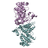
| Title | Crystal structure of a ternary complex of the transcriptional repressor Gal80p (Gal80S0 [G301R]) and the acidic activation domain of Gal4p (aa 854-874) from Saccharomyces cerevisiae with NAD | ||||||
 Components Components |
| ||||||
 Keywords Keywords |  TRANSCRIPTION / Eukaryotic transcription complex / NAD / Rossmann fold / Acetylation / Carbohydrate metabolism / DNA-binding / Galactose metabolism / Repressor / Transcription regulation / Activator / Metal-binding / Nucleus / Phosphoprotein / Zinc TRANSCRIPTION / Eukaryotic transcription complex / NAD / Rossmann fold / Acetylation / Carbohydrate metabolism / DNA-binding / Galactose metabolism / Repressor / Transcription regulation / Activator / Metal-binding / Nucleus / Phosphoprotein / Zinc | ||||||
| Function / homology |  Function and homology informationregulation of transcription from RNA polymerase II promoter by galactose / kinase inhibitor activity / galactose metabolic process / negative regulation of phosphorylation / positive regulation of transcription from RNA polymerase II promoter by galactose / transcription repressor complex / DNA-binding transcription activator activity, RNA polymerase II-specific / RNA polymerase II-specific DNA-binding transcription factor binding / transcription regulator complex / DNA-binding transcription factor activity, RNA polymerase II-specific ...regulation of transcription from RNA polymerase II promoter by galactose / kinase inhibitor activity / galactose metabolic process / negative regulation of phosphorylation / positive regulation of transcription from RNA polymerase II promoter by galactose / transcription repressor complex / DNA-binding transcription activator activity, RNA polymerase II-specific / RNA polymerase II-specific DNA-binding transcription factor binding / transcription regulator complex / DNA-binding transcription factor activity, RNA polymerase II-specific / RNA polymerase II cis-regulatory region sequence-specific DNA binding / nucleotide binding / DNA-templated transcription / negative regulation of transcription by RNA polymerase II / DNA binding / zinc ion binding / identical protein binding / nucleus / cytoplasm Function and homology informationregulation of transcription from RNA polymerase II promoter by galactose / kinase inhibitor activity / galactose metabolic process / negative regulation of phosphorylation / positive regulation of transcription from RNA polymerase II promoter by galactose / transcription repressor complex / DNA-binding transcription activator activity, RNA polymerase II-specific / RNA polymerase II-specific DNA-binding transcription factor binding / transcription regulator complex / DNA-binding transcription factor activity, RNA polymerase II-specific ...regulation of transcription from RNA polymerase II promoter by galactose / kinase inhibitor activity / galactose metabolic process / negative regulation of phosphorylation / positive regulation of transcription from RNA polymerase II promoter by galactose / transcription repressor complex / DNA-binding transcription activator activity, RNA polymerase II-specific / RNA polymerase II-specific DNA-binding transcription factor binding / transcription regulator complex / DNA-binding transcription factor activity, RNA polymerase II-specific / RNA polymerase II cis-regulatory region sequence-specific DNA binding / nucleotide binding / DNA-templated transcription / negative regulation of transcription by RNA polymerase II / DNA binding / zinc ion binding / identical protein binding / nucleus / cytoplasmSimilarity search - Function | ||||||
| Biological species |  Saccharomyces cerevisiae (brewer's yeast) Saccharomyces cerevisiae (brewer's yeast) | ||||||
| Method | X-RAY DIFFRACTION / SYNCHROTRON / MOLECULAR REPLACEMENT / Resolution: 2.7 Å | ||||||
 Authors Authors | Kumar, P.R. / Joshua-Tor, L. | ||||||
 Citation Citation | Journal: Science / Year: 2008 Title: NADP regulates the yeast GAL induction system. Authors: Kumar, P.R. / Yu, Y. / Sternglanz, R. / Johnston, S.A. / Joshua-Tor, L. | ||||||
| History |
|
- Structure visualization
| Structure viewer | Molecule: MolmilJmol/JSmol |
|---|
- Downloads & links
-Download
| PDBx/mmCIF format | 3bts.cif.gz | 175.2 KB | Display | PDBx/mmCIF format |
|---|---|---|---|---|
| PDB format | pdb3bts.ent.gz | 138.8 KB | Display | PDB format |
| PDBx/mmJSON format | 3bts.json.gz | Tree view | PDBx/mmJSON format | |
| Others |  Other downloads Other downloads |
-Validation report
| Arichive directory | https://data.pdbj.org/pub/pdb/validation_reports/bt/3btsftp://data.pdbj.org/pub/pdb/validation_reports/bt/3bts | HTTPS FTP |
|---|
-Related structure data
| Related structure data |  3btuSC  3btvC S: Starting model for refinement C: citing same article ( |
|---|---|
| Similar structure data |


-Links
 PDBj
PDBj- Assembly
| Deposited unit | 
| ||||||||
|---|---|---|---|---|---|---|---|---|---|
| 1 |
| ||||||||
| Unit cell |
|
-Components
| #1: Protein | Mass: 48754.434 Da / Num. of mol.: 2 / Mutation: G301R Source method: isolated from a genetically manipulated source Source: (gene. exp.) Saccharomyces cerevisiae (brewer's yeast)Gene: GAL80 / Plasmid: PET28a / Production host:  Escherichia coli (E. coli) / Strain (production host): BL21(DE3)-RIPL / References: UniProt: P04387 Escherichia coli (E. coli) / Strain (production host): BL21(DE3)-RIPL / References: UniProt: P04387#2: Protein/peptide | Mass: 2507.616 Da / Num. of mol.: 2 / Fragment: S. cerevisiae Gal4p peptide; UNP residues 854-874 / Source method: obtained synthetically / Details: S.cerevisiae Gal4(854-874) was made synthetically / References: UniProt: P04386 #3: Chemical | Nicotinamide adenine dinucleotide  Mass: 663.425 Da / Num. of mol.: 2 / Source method: obtained synthetically / Formula: C21H27N7O14P2 / Comment: NAD*YM Mass: 663.425 Da / Num. of mol.: 2 / Source method: obtained synthetically / Formula: C21H27N7O14P2 / Comment: NAD*YM#4: Water | ChemComp-HOH / | Water Mass: 18.015 Da / Num. of mol.: 127 / Source method: isolated from a natural source / Formula: H2O Mass: 18.015 Da / Num. of mol.: 127 / Source method: isolated from a natural source / Formula: H2O |
|---|
-Experimental details
-Experiment
| Experiment | Method: X-RAY DIFFRACTION / Number of used crystals: 1 |
|---|
- Sample preparation
| Crystal | Density Matthews: 2.37 Å3/Da / Density % sol: 48.19 % |
|---|---|
| Crystal grow | Temperature: 290 K / Method: vapor diffusion, hanging drop / pH: 8 Details: 20% PEG 3350, 0.15M Sodium fluoride, followed by soaking in NAD to a final concentration of 5mM, pH 8.0, VAPOR DIFFUSION, HANGING DROP, temperature 290K |
-Data collection
| Diffraction | Mean temperature: 100 K |
|---|---|
| Diffraction source | Source: SYNCHROTRON / Site: NSLS  / Beamline: X29A / Wavelength: 0.9792 Å / Beamline: X29A / Wavelength: 0.9792 Å |
| Detector | Type: ADSC QUANTUM 315 / Detector: CCD / Details: monochromator |
| Radiation | Protocol: SINGLE WAVELENGTH / Monochromatic (M) / Laue (L): M / Scattering type: x-ray |
| Radiation wavelength | Wavelength: 0.9792 Å / Relative weight: 1 |
| Reflection | Resolution: 2.7→50 Å / Num. all: 27466 / Num. obs: 27319 / % possible obs: 99.5 % / Observed criterion σ(I): -3 / Redundancy: 6.5 % / Biso Wilson estimate: 58.854 Å2 / Rmerge(I) obs: 0.091 / Net I/σ(I): 19.1 |
| Reflection shell | Resolution: 2.7→2.8 Å / Redundancy: 4.1 % / Rmerge(I) obs: 0.48 / Mean I/σ(I) obs: 1.87 / % possible all: 95.6 |
- Processing
| Software |
| |||||||||||||||||||||||||||||||||||||||||||||||||||||||||||||||||||||||||||||||||||||||||||||||||||||||||||||||||||||||||||||||||||||||||||||||||||||||||||||||||||||||||||||||||||||||||||||||||||||||||||||||||||||||||||||||||||||||||||||||||||||||||||||||||||||||||||||||||||
|---|---|---|---|---|---|---|---|---|---|---|---|---|---|---|---|---|---|---|---|---|---|---|---|---|---|---|---|---|---|---|---|---|---|---|---|---|---|---|---|---|---|---|---|---|---|---|---|---|---|---|---|---|---|---|---|---|---|---|---|---|---|---|---|---|---|---|---|---|---|---|---|---|---|---|---|---|---|---|---|---|---|---|---|---|---|---|---|---|---|---|---|---|---|---|---|---|---|---|---|---|---|---|---|---|---|---|---|---|---|---|---|---|---|---|---|---|---|---|---|---|---|---|---|---|---|---|---|---|---|---|---|---|---|---|---|---|---|---|---|---|---|---|---|---|---|---|---|---|---|---|---|---|---|---|---|---|---|---|---|---|---|---|---|---|---|---|---|---|---|---|---|---|---|---|---|---|---|---|---|---|---|---|---|---|---|---|---|---|---|---|---|---|---|---|---|---|---|---|---|---|---|---|---|---|---|---|---|---|---|---|---|---|---|---|---|---|---|---|---|---|---|---|---|---|---|---|---|---|---|---|---|---|---|---|---|---|---|---|---|---|---|---|---|---|---|---|---|---|---|---|---|---|---|---|---|---|---|---|---|---|---|---|---|---|---|---|---|---|---|---|---|---|---|---|---|---|
| Refinement | Method to determine structure: MOLECULAR REPLACEMENT Starting model: PDB ENTRY 3BTU Resolution: 2.7→46.57 Å / Cor.coef. Fo:Fc: 0.909 / Cor.coef. Fo:Fc free: 0.884 / SU B: 29.914 / SU ML: 0.287 / Cross valid method: THROUGHOUT / σ(F): 0 / ESU R: 1.398 / ESU R Free: 0.374 / Stereochemistry target values: MAXIMUM LIKELIHOOD / Details: HYDROGENS HAVE BEEN ADDED IN THE RIDING POSITIONS
| |||||||||||||||||||||||||||||||||||||||||||||||||||||||||||||||||||||||||||||||||||||||||||||||||||||||||||||||||||||||||||||||||||||||||||||||||||||||||||||||||||||||||||||||||||||||||||||||||||||||||||||||||||||||||||||||||||||||||||||||||||||||||||||||||||||||||||||||||||
| Solvent computation | Ion probe radii: 0.8 Å / Shrinkage radii: 0.8 Å / VDW probe radii: 1.4 Å / Solvent model: BABINET MODEL WITH MASK | |||||||||||||||||||||||||||||||||||||||||||||||||||||||||||||||||||||||||||||||||||||||||||||||||||||||||||||||||||||||||||||||||||||||||||||||||||||||||||||||||||||||||||||||||||||||||||||||||||||||||||||||||||||||||||||||||||||||||||||||||||||||||||||||||||||||||||||||||||
| Refine analyze | Luzzati coordinate error obs: 0.468 Å | |||||||||||||||||||||||||||||||||||||||||||||||||||||||||||||||||||||||||||||||||||||||||||||||||||||||||||||||||||||||||||||||||||||||||||||||||||||||||||||||||||||||||||||||||||||||||||||||||||||||||||||||||||||||||||||||||||||||||||||||||||||||||||||||||||||||||||||||||||
| Refinement step | Cycle: LAST / Resolution: 2.7→46.57 Å
| |||||||||||||||||||||||||||||||||||||||||||||||||||||||||||||||||||||||||||||||||||||||||||||||||||||||||||||||||||||||||||||||||||||||||||||||||||||||||||||||||||||||||||||||||||||||||||||||||||||||||||||||||||||||||||||||||||||||||||||||||||||||||||||||||||||||||||||||||||
| LS refinement shell | Resolution: 2.7→2.79 Å / Total num. of bins used: 10
| |||||||||||||||||||||||||||||||||||||||||||||||||||||||||||||||||||||||||||||||||||||||||||||||||||||||||||||||||||||||||||||||||||||||||||||||||||||||||||||||||||||||||||||||||||||||||||||||||||||||||||||||||||||||||||||||||||||||||||||||||||||||||||||||||||||||||||||||||||
| Refinement TLS params. | Method: refined / Refine-ID: X-RAY DIFFRACTION
| |||||||||||||||||||||||||||||||||||||||||||||||||||||||||||||||||||||||||||||||||||||||||||||||||||||||||||||||||||||||||||||||||||||||||||||||||||||||||||||||||||||||||||||||||||||||||||||||||||||||||||||||||||||||||||||||||||||||||||||||||||||||||||||||||||||||||||||||||||
| Refinement TLS group |
|
