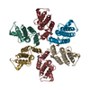
Deposited unit
A: Gag protein
B: Gag protein
C: Gag protein
D: Gag protein
E: Gag protein
F: Gag protein
G: Gag protein
H: Gag protein
I: Gag protein
J: Gag protein
K: Gag protein
L: Gag protein
M: Gag protein
N: Gag protein
O: Gag protein
P: Gag protein
Q: Gag protein
R: Gag protein
S: Gag protein
T: Gag protein
U: Gag protein
V: Gag protein
X: Gag protein
Y: Gag protein
hetero molecules Summary Component details
Theoretical mass Number of molelcules Total (without water) 384,310 37 Polymers 383,464 24 Non-polymers 845 13 Water 5,206 289
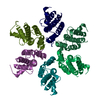
1
A: Gag protein
B: Gag protein
C: Gag protein
D: Gag protein
E: Gag protein
F: Gag protein
hetero molecules Summary Component details Symmetry operations
Theoretical mass Number of molelcules Total (without water) 96,319 13 Polymers 95,866 6 Non-polymers 453 7 Water 108 6
Type Name Symmetry operation Number identity operation 1_555 x,y,z 1
2
G: Gag protein
H: Gag protein
I: Gag protein
J: Gag protein
K: Gag protein
L: Gag protein
hetero molecules Summary Component details Symmetry operations
Theoretical mass Number of molelcules Total (without water) 96,259 12 Polymers 95,866 6 Non-polymers 393 6 Water 108 6
Type Name Symmetry operation Number identity operation 1_555 x,y,z 1
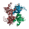
3
M: Gag protein
N: Gag protein
O: Gag protein
P: Gag protein
Q: Gag protein
R: Gag protein Summary Component details Symmetry operations
Theoretical mass Number of molelcules Total (without water) 95,866 6 Polymers 95,866 6 Non-polymers 0 0 Water 108 6
Type Name Symmetry operation Number identity operation 1_555 x,y,z 1
4
S: Gag protein
T: Gag protein
U: Gag protein
V: Gag protein
X: Gag protein
Y: Gag protein Summary Component details Symmetry operations
Theoretical mass Number of molelcules Total (without water) 95,866 6 Polymers 95,866 6 Non-polymers 0 0 Water 108 6
Type Name Symmetry operation Number identity operation 1_555 x,y,z 1
Unit cell Length a, b, c (Å) 86.264, 86.832, 152.360 Angle α, β, γ (deg.) 89.120, 90.040, 60.260 Int Tables number 1 Space group name H-M P1
Noncrystallographic symmetry (NCS) NCS domain Show large table (3 x 72) Hide large table ID Ens-ID Details 1 1 A2 1 B3 1 C4 1 D5 1 E6 1 F7 1 G8 1 H9 1 I10 1 J11 1 K12 1 L13 1 M14 1 N15 1 O16 1 P17 1 Q18 1 R19 1 S20 1 T21 1 U22 1 V23 1 X24 1 Y25 1 A26 1 B27 1 C28 1 D29 1 E30 1 F31 1 G32 1 H33 1 I34 1 J35 1 K36 1 L37 1 M38 1 N39 1 O40 1 P41 1 Q42 1 R43 1 S44 1 T45 1 U46 1 V47 1 X48 1 Y49 1 A50 1 B51 1 C52 1 D53 1 E54 1 F55 1 G56 1 H57 1 I58 1 J59 1 K60 1 L61 1 M62 1
 Movie
Movie Controller


 Open data
 Basic information
 Components
 HIV-1 protease
  Keywords
 Function and homology information
 Authors
 Citation
 Structure visualization
 Downloads & links
 Other downloads
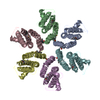


 PDBj
PDBj
 Assembly