 Movie
Movie Controller

[English] 日本語
 Yorodumi
- PDB-1u7k: Structure of a hexameric N-terminal domain from murine leukemia v... -
+ Open data

- Basic information
| Entry | Database: PDB / ID: 1u7k | ||||||
|---|---|---|---|---|---|---|---|
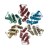
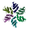
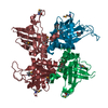
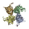
| Title | Structure of a hexameric N-terminal domain from murine leukemia virus capsid | ||||||
 Components Components | Gag polyprotein Group-specific antigen Group-specific antigen | ||||||
 Keywords Keywords | VIRAL PROTEIN / capsid | ||||||
| Function / homology |  Function and homology information Function and homology informationviral budding via host ESCRT complex / host multivesicular body / viral nucleocapsid / structural constituent of virion / host cell plasma membrane / RNA binding / zinc ion binding / membraneSimilarity search - Function | ||||||
| Biological species | AKR murine leukemia virus | ||||||
| Method | X-RAY DIFFRACTION / SYNCHROTRON / MAD / Resolution: 1.85 Å | ||||||
 Authors Authors | Mortuza, G.B. / Haire, L.F. / Stevens, A. / Smerdon, S.J. / Stoye, J.P. / Taylor, I.A. | ||||||
 Citation Citation | Journal: Nature / Year: 2004 Title: High-resolution structure of a retroviral capsid hexameric amino-terminal domain. Authors: Mortuza, G.B. / Haire, L.F. / Stevens, A. / Smerdon, S.J. / Stoye, J.P. / Taylor, I.A. | ||||||
| History |
|
- Structure visualization
| Structure viewer | Molecule: MolmilJmol/JSmol |
|---|
- Downloads & links
-Download
| PDBx/mmCIF format | 1u7k.cif.gz | 165.1 KB | Display | PDBx/mmCIF format |
|---|---|---|---|---|
| PDB format | pdb1u7k.ent.gz | 139.6 KB | Display | PDB format |
| PDBx/mmJSON format | 1u7k.json.gz | Tree view | PDBx/mmJSON format | |
| Others |  Other downloads Other downloads |
-Validation report
| Arichive directory | https://data.pdbj.org/pub/pdb/validation_reports/u7/1u7kftp://data.pdbj.org/pub/pdb/validation_reports/u7/1u7k | HTTPS FTP |
|---|
-Related structure data
| Similar structure data |
|---|


-Links
 PDBj
PDBj- Assembly
| Deposited unit | 
| ||||||||
|---|---|---|---|---|---|---|---|---|---|
| 1 |
| ||||||||
| Unit cell |
|
-Components
| #1: Protein | Group-specific antigen Mass: 15004.415 Da / Num. of mol.: 6 / Fragment: n-terminal domain (residues 215-345) Source method: isolated from a genetically manipulated source Source: (gene. exp.)  AKR (endogenous) murine leukemia virus / Genus: Gammaretrovirus / Species: Murine leukemia virus / Gene: GAG / Plasmid: pET22b / Species (production host): Escherichia coli / Production host: AKR (endogenous) murine leukemia virus / Genus: Gammaretrovirus / Species: Murine leukemia virus / Gene: GAG / Plasmid: pET22b / Species (production host): Escherichia coli / Production host:  Escherichia coli BL21(DE3) (bacteria) / Strain (production host): bl21(DE3) / References: UniProt: P03336 Escherichia coli BL21(DE3) (bacteria) / Strain (production host): bl21(DE3) / References: UniProt: P03336#2: Water | ChemComp-HOH / | Water Mass: 18.015 Da / Num. of mol.: 431 / Source method: isolated from a natural source / Formula: H2O Mass: 18.015 Da / Num. of mol.: 431 / Source method: isolated from a natural source / Formula: H2O |
|---|
-Experimental details
-Experiment
| Experiment | Method: X-RAY DIFFRACTION / Number of used crystals: 1 |
|---|
- Sample preparation
| Crystal | Density Matthews: 2.81 Å3/Da / Density % sol: 56.23 % |
|---|
-Data collection
| Diffraction | Mean temperature: 100 K | ||||||||||||
|---|---|---|---|---|---|---|---|---|---|---|---|---|---|
| Diffraction source | Source: SYNCHROTRON / Site: SRS  / Beamline: PX14.2 / Wavelength: 0.9779, 0.970, 0.9783 / Beamline: PX14.2 / Wavelength: 0.9779, 0.970, 0.9783 | ||||||||||||
| Detector | Type: ADSC QUANTUM 4 / Detector: CCD / Date: Feb 1, 2004 | ||||||||||||
| Radiation | Protocol: MAD / Monochromatic (M) / Laue (L): M / Scattering type: x-ray | ||||||||||||
| Radiation wavelength |
| ||||||||||||
| Reflection | Resolution: 1.85→20 Å / Num. all: 85390 / Num. obs: 85291 / % possible obs: 100 % / Observed criterion σ(F): 0 / Observed criterion σ(I): 2 / Rmerge(I) obs: 0.081 | ||||||||||||
| Reflection shell | Resolution: 1.9→1.98 Å / Rmerge(I) obs: 0.44 / % possible all: 100 |
- Processing
| Software |
| ||||||||||||||||||||||||||||||||||||||||||||||||||||||||||||||||||||||||||||||||||||||||||||||||||||||||||||||||||||||||||||||||||||||||||||||||||||||||||||||||
|---|---|---|---|---|---|---|---|---|---|---|---|---|---|---|---|---|---|---|---|---|---|---|---|---|---|---|---|---|---|---|---|---|---|---|---|---|---|---|---|---|---|---|---|---|---|---|---|---|---|---|---|---|---|---|---|---|---|---|---|---|---|---|---|---|---|---|---|---|---|---|---|---|---|---|---|---|---|---|---|---|---|---|---|---|---|---|---|---|---|---|---|---|---|---|---|---|---|---|---|---|---|---|---|---|---|---|---|---|---|---|---|---|---|---|---|---|---|---|---|---|---|---|---|---|---|---|---|---|---|---|---|---|---|---|---|---|---|---|---|---|---|---|---|---|---|---|---|---|---|---|---|---|---|---|---|---|---|---|---|---|---|
| Refinement | Method to determine structure: MAD / Resolution: 1.85→12 Å / Cor.coef. Fo:Fc: 0.936 / Cor.coef. Fo:Fc free: 0.918 / SU B: 3.11 / SU ML: 0.096 / Cross valid method: THROUGHOUT / σ(F): 0 / ESU R: 0.14 / ESU R Free: 0.132 / Stereochemistry target values: MAXIMUM LIKELIHOOD
| ||||||||||||||||||||||||||||||||||||||||||||||||||||||||||||||||||||||||||||||||||||||||||||||||||||||||||||||||||||||||||||||||||||||||||||||||||||||||||||||||
| Solvent computation | Ion probe radii: 0.8 Å / Shrinkage radii: 0.8 Å / VDW probe radii: 1.4 Å / Solvent model: BABINET MODEL WITH MASK | ||||||||||||||||||||||||||||||||||||||||||||||||||||||||||||||||||||||||||||||||||||||||||||||||||||||||||||||||||||||||||||||||||||||||||||||||||||||||||||||||
| Displacement parameters | Biso mean: 22.068 Å2
| ||||||||||||||||||||||||||||||||||||||||||||||||||||||||||||||||||||||||||||||||||||||||||||||||||||||||||||||||||||||||||||||||||||||||||||||||||||||||||||||||
| Refinement step | Cycle: LAST / Resolution: 1.85→12 Å
| ||||||||||||||||||||||||||||||||||||||||||||||||||||||||||||||||||||||||||||||||||||||||||||||||||||||||||||||||||||||||||||||||||||||||||||||||||||||||||||||||
| Refine LS restraints |
| ||||||||||||||||||||||||||||||||||||||||||||||||||||||||||||||||||||||||||||||||||||||||||||||||||||||||||||||||||||||||||||||||||||||||||||||||||||||||||||||||
| LS refinement shell | Resolution: 1.85→1.897 Å / Total num. of bins used: 20 /
|
