 Movie
Movie Controller

[English] 日本語
 Yorodumi
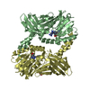
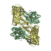
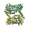
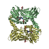
- PDB-3b3g: The 2.4 A crystal structure of the apo catalytic domain of coacti... -
+ Open data

- Basic information
| Entry | Database: PDB / ID: 3b3g | ||||||
|---|---|---|---|---|---|---|---|
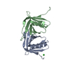
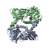
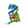
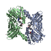
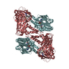
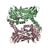
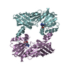
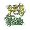
| Title | The 2.4 A crystal structure of the apo catalytic domain of coactivator-associated arginine methyl transferase I(CARM1,140-480). | ||||||
 Components Components | Histone-arginine methyltransferase CARM1 | ||||||
 Keywords Keywords |  TRANSFERASE / PROTEIN ARGININE METHYLTRANSFERASE / CATALYTIC DOMAIN / Chromatin regulator / mRNA processing / mRNA splicing / Nucleus / S-adenosyl-L-methionine / Transcription / Transcription regulation TRANSFERASE / PROTEIN ARGININE METHYLTRANSFERASE / CATALYTIC DOMAIN / Chromatin regulator / mRNA processing / mRNA splicing / Nucleus / S-adenosyl-L-methionine / Transcription / Transcription regulation | ||||||
| Function / homology |  Function and homology information Function and homology informationRMTs methylate histone arginines / Estrogen-dependent gene expression / Regulation of lipid metabolism by PPARalpha / histone H3R26 methyltransferase activity / Cytoprotection by HMOX1 / regulation of growth plate cartilage chondrocyte proliferation / histone H3R17 methyltransferase activity / endochondral bone morphogenesis / histone H3R2 methyltransferase activity / negative regulation of dendrite development ...RMTs methylate histone arginines / Estrogen-dependent gene expression / Regulation of lipid metabolism by PPARalpha / histone H3R26 methyltransferase activity / Cytoprotection by HMOX1 / regulation of growth plate cartilage chondrocyte proliferation / histone H3R17 methyltransferase activity / endochondral bone morphogenesis / histone H3R2 methyltransferase activity / negative regulation of dendrite development / type I protein arginine methyltransferase / protein-arginine omega-N asymmetric methyltransferase activity / protein methyltransferase activity / histone arginine N-methyltransferase activity / regulation of intracellular estrogen receptor signaling pathway / replication fork reversal / protein-arginine N-methyltransferase activity / positive regulation of epithelial cell apoptotic process / histone methyltransferase activity / positive regulation of transcription by RNA polymerase I / nuclear replication fork / positive regulation of fat cell differentiation / intracellular estrogen receptor signaling pathway / intracellular steroid hormone receptor signaling pathway / response to cAMP / protein localization to chromatin / RNA splicing / nuclear receptor coactivator activity / lysine-acetylated histone binding / mRNA processing / RNA polymerase II transcription regulator complex / methylation / DNA-binding transcription factor binding / cell population proliferation / transcription coactivator activity / transcription cis-regulatory region binding / positive regulation of cell population proliferation / regulation of DNA-templated transcription / protein homodimerization activity / positive regulation of transcription by RNA polymerase II / protein-containing complex / nucleoplasm / nucleus / cytosol / cytoplasmSimilarity search - Function | ||||||
| Biological species |  Rattus norvegicus (Norway rat) Rattus norvegicus (Norway rat) | ||||||
| Method | X-RAY DIFFRACTION / SYNCHROTRON / MOLECULAR REPLACEMENT / Resolution: 2.4 Å | ||||||
 Authors Authors | Troffer-Charlier, N. / Cura, V. / Hassenboehler, P. / Moras, D. / Cavarelli, J. | ||||||
 Citation Citation | Journal: Embo J. / Year: 2007 Title: Functional insights from structures of coactivator-associated arginine methyltransferase 1 domains. Authors: Troffer-Charlier, N. / Cura, V. / Hassenboehler, P. / Moras, D. / Cavarelli, J. #1: Journal: Acta Crystallogr.,Sect.F / Year: 2007 Title: Expression, purification, crystallization and preliminary crystallographic study of isolated modules of the mouse coactivator-associated arginine methyltransferase 1. Authors: Troffer-Charlier, N. / Cura, V. / Hassenboehler, P. / Moras, D. / Cavarelli, J. | ||||||
| History |
|
- Structure visualization
| Structure viewer | Molecule: MolmilJmol/JSmol |
|---|
- Downloads & links
-Download
| PDBx/mmCIF format | 3b3g.cif.gz | 137.5 KB | Display | PDBx/mmCIF format |
|---|---|---|---|---|
| PDB format | pdb3b3g.ent.gz | 108.2 KB | Display | PDB format |
| PDBx/mmJSON format | 3b3g.json.gz | Tree view | PDBx/mmJSON format | |
| Others |  Other downloads Other downloads |
-Validation report
| Arichive directory | https://data.pdbj.org/pub/pdb/validation_reports/b3/3b3gftp://data.pdbj.org/pub/pdb/validation_reports/b3/3b3g | HTTPS FTP |
|---|
-Related structure data

-Links
 PDBj
PDBj- Assembly
| Deposited unit | 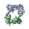
| ||||||||
|---|---|---|---|---|---|---|---|---|---|
| 1 |
| ||||||||
| Unit cell |
|
-Components
| #1: Protein | Mass: 38774.160 Da / Num. of mol.: 2 / Fragment: Catalytic Domain Source method: isolated from a genetically manipulated source Source: (gene. exp.) Rattus norvegicus (Norway rat) / Gene: Carm1, Prmt4 / Production host:  unidentified baculovirus / Strain (production host): SF9 unidentified baculovirus / Strain (production host): SF9References: UniProt: Q4AE70, EC: 2.1.1.125, Transferases; Transferring one-carbon groups; Methyltransferases#2: Water | ChemComp-HOH / | Water Mass: 18.015 Da / Num. of mol.: 57 / Source method: isolated from a natural source / Formula: H2O Mass: 18.015 Da / Num. of mol.: 57 / Source method: isolated from a natural source / Formula: H2O |
|---|
-Experimental details
-Experiment
| Experiment | Method: X-RAY DIFFRACTION / Number of used crystals: 1 |
|---|
- Sample preparation
| Crystal | Density Matthews: 2.42 Å3/Da / Density % sol: 49.13 % |
|---|---|
| Crystal grow | Temperature: 297 K / Method: vapor diffusion, hanging drop / pH: 7 Details: 19% PEG 3350, 0.15M sodium malate, pH 7.0, VAPOR DIFFUSION, HANGING DROP, temperature 297K |
-Data collection
| Diffraction | Mean temperature: 100 K |
|---|---|
| Diffraction source | Source: SYNCHROTRON / Site: ESRF  / Beamline: ID23-1 / Beamline: ID23-1 |
| Detector | Type: ADSC QUANTUM 315 / Detector: CCD / Date: May 15, 2007 |
| Radiation | Protocol: SINGLE WAVELENGTH / Monochromatic (M) / Laue (L): M / Scattering type: x-ray |
| Radiation wavelength | Relative weight: 1 |
| Reflection | Resolution: 2.4→29.49 Å / Num. all: 30000 / Num. obs: 28039 / % possible obs: 98.9 % / Observed criterion σ(F): 0 / Observed criterion σ(I): 0 / Redundancy: 4.2 % / Biso Wilson estimate: 49.9 Å2 / Rmerge(I) obs: 0.045 / Rsym value: 0.045 / Net I/σ(I): 20.8 |
| Reflection shell | Resolution: 2.4→2.49 Å / Redundancy: 4 % / Rmerge(I) obs: 0.177 / Mean I/σ(I) obs: 6.8 / Num. unique all: 2791 / Rsym value: 0.177 / % possible all: 98.9 |
- Processing
| Software |
| ||||||||||||||||||||||||||||||||||||||||||||||||||||||||||||||||||||||||||||||||||||||||||||||||||||||||||||||||||||||||||||||||||||||||||||||||||||||||||||||||||||||||||
|---|---|---|---|---|---|---|---|---|---|---|---|---|---|---|---|---|---|---|---|---|---|---|---|---|---|---|---|---|---|---|---|---|---|---|---|---|---|---|---|---|---|---|---|---|---|---|---|---|---|---|---|---|---|---|---|---|---|---|---|---|---|---|---|---|---|---|---|---|---|---|---|---|---|---|---|---|---|---|---|---|---|---|---|---|---|---|---|---|---|---|---|---|---|---|---|---|---|---|---|---|---|---|---|---|---|---|---|---|---|---|---|---|---|---|---|---|---|---|---|---|---|---|---|---|---|---|---|---|---|---|---|---|---|---|---|---|---|---|---|---|---|---|---|---|---|---|---|---|---|---|---|---|---|---|---|---|---|---|---|---|---|---|---|---|---|---|---|---|---|---|---|
| Refinement | Method to determine structure: MOLECULAR REPLACEMENT / Resolution: 2.4→29.49 Å / Cor.coef. Fo:Fc: 0.943 / Cor.coef. Fo:Fc free: 0.902 / SU B: 17.845 / SU ML: 0.197 / TLS residual ADP flag: LIKELY RESIDUAL / Cross valid method: THROUGHOUT / σ(F): 0 / σ(I): 0 / ESU R: 0.432 / ESU R Free: 0.281 / Stereochemistry target values: MAXIMUM LIKELIHOOD / Details: HYDROGENS HAVE BEEN ADDED IN THE RIDING POSITIONS
| ||||||||||||||||||||||||||||||||||||||||||||||||||||||||||||||||||||||||||||||||||||||||||||||||||||||||||||||||||||||||||||||||||||||||||||||||||||||||||||||||||||||||||
| Solvent computation | Ion probe radii: 0.8 Å / Shrinkage radii: 0.8 Å / VDW probe radii: 1.2 Å / Solvent model: MASK | ||||||||||||||||||||||||||||||||||||||||||||||||||||||||||||||||||||||||||||||||||||||||||||||||||||||||||||||||||||||||||||||||||||||||||||||||||||||||||||||||||||||||||
| Displacement parameters | Biso mean: 44.711 Å2
| ||||||||||||||||||||||||||||||||||||||||||||||||||||||||||||||||||||||||||||||||||||||||||||||||||||||||||||||||||||||||||||||||||||||||||||||||||||||||||||||||||||||||||
| Refinement step | Cycle: LAST / Resolution: 2.4→29.49 Å
| ||||||||||||||||||||||||||||||||||||||||||||||||||||||||||||||||||||||||||||||||||||||||||||||||||||||||||||||||||||||||||||||||||||||||||||||||||||||||||||||||||||||||||
| Refine LS restraints |
| ||||||||||||||||||||||||||||||||||||||||||||||||||||||||||||||||||||||||||||||||||||||||||||||||||||||||||||||||||||||||||||||||||||||||||||||||||||||||||||||||||||||||||
| LS refinement shell | Resolution: 2.4→2.462 Å / Total num. of bins used: 20
| ||||||||||||||||||||||||||||||||||||||||||||||||||||||||||||||||||||||||||||||||||||||||||||||||||||||||||||||||||||||||||||||||||||||||||||||||||||||||||||||||||||||||||
| Refinement TLS params. | Method: refined / Refine-ID: X-RAY DIFFRACTION
| ||||||||||||||||||||||||||||||||||||||||||||||||||||||||||||||||||||||||||||||||||||||||||||||||||||||||||||||||||||||||||||||||||||||||||||||||||||||||||||||||||||||||||
| Refinement TLS group |
|
