| 登録情報 | データベース: PDB / ID: 3atp
|
|---|
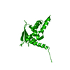
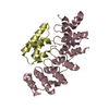
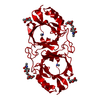
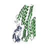
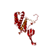
| タイトル | Structure of the ligand binding domain of the bacterial serine chemoreceptor Tsr with ligand |
|---|
 要素 要素 | Methyl-accepting chemotaxis protein I |
|---|
 キーワード キーワード |  SIGNALING PROTEIN / Chemoreceptor (化学受容器) / serine biniding / membrane (生体膜) SIGNALING PROTEIN / Chemoreceptor (化学受容器) / serine biniding / membrane (生体膜) |
|---|
| 機能・相同性 |  機能・相同性情報 機能・相同性情報
regulation of protein histidine kinase activity / detection of chemical stimulus / methyl accepting chemotaxis protein complex / regulation of bacterial-type flagellum-dependent cell motility / regulation of chemotaxis / signal complex assembly / receptor clustering / 運動性 / cellular response to amino acid stimulus / transmembrane signaling receptor activity ...regulation of protein histidine kinase activity / detection of chemical stimulus / methyl accepting chemotaxis protein complex / regulation of bacterial-type flagellum-dependent cell motility / regulation of chemotaxis / signal complex assembly / receptor clustering / 運動性 / cellular response to amino acid stimulus / transmembrane signaling receptor activity / 走化性 / シグナル伝達 / identical protein binding / 細胞膜類似検索 - 分子機能 Aspartate receptor, ligand-binding domain / Methyl-accepting chemotaxis protein, four helix bundle domain superfamily / Homologues of the ligand binding domain of Tar / Chemotaxis methyl-accepting receptor, methyl-accepting site / Bacterial chemotaxis sensory transducers signature. / Chemotaxis methyl-accepting receptor Tar-related, ligand-binding / Tar ligand binding domain homologue / Chemotaxis methyl-accepting receptor / Methyl-accepting chemotaxis protein (MCP) signalling domain / Methyl-accepting chemotaxis protein (MCP) signalling domain ...Aspartate receptor, ligand-binding domain / Methyl-accepting chemotaxis protein, four helix bundle domain superfamily / Homologues of the ligand binding domain of Tar / Chemotaxis methyl-accepting receptor, methyl-accepting site / Bacterial chemotaxis sensory transducers signature. / Chemotaxis methyl-accepting receptor Tar-related, ligand-binding / Tar ligand binding domain homologue / Chemotaxis methyl-accepting receptor / Methyl-accepting chemotaxis protein (MCP) signalling domain / Methyl-accepting chemotaxis protein (MCP) signalling domain / Bacterial chemotaxis sensory transducers domain profile. / Methyl-accepting chemotaxis-like domains (chemotaxis sensory transducer). / HAMP domain / HAMP (Histidine kinases, Adenylyl cyclases, Methyl binding proteins, Phosphatases) domain / HAMP domain profile. / HAMP domain / Four Helix Bundle (Hemerythrin (Met), subunit A) / Up-down Bundle / Mainly Alpha類似検索 - ドメイン・相同性 |
|---|
| 生物種 |   Escherichia coli (大腸菌) Escherichia coli (大腸菌) |
|---|
| 手法 | X線回折 / シンクロトロン / 分子置換 / 解像度: 2.5 Å |
|---|
 データ登録者 データ登録者 | Tajima, H. / Sakuma, M. / Homma, K. / Kawagishi, I. / Imada, K. |
|---|
 引用 引用 | ジャーナル: J.Biol.Chem. / 年: 2011
タイトル: Ligand specificity determined by differentially arranged common ligand-binding residues in bacterial amino acid chemoreceptors Tsr and Tar.
著者: Tajima, H. / Imada, K. / Sakuma, M. / Hattori, F. / Nara, T. / Kamo, N. / Homma, M. / Kawagishi, I. |
|---|
| 履歴 | | 登録 | 2011年1月7日 | 登録サイト: PDBJ / 処理サイト: PDBJ |
|---|
| 改定 1.0 | 2011年10月19日 | Provider: repository / タイプ: Initial release |
|---|
| 改定 1.1 | 2013年6月19日 | Group: Database references |
|---|
| 改定 1.2 | 2024年3月13日 | Group: Data collection / Database references / Derived calculations
カテゴリ: chem_comp_atom / chem_comp_bond ...chem_comp_atom / chem_comp_bond / database_2 / struct_ref_seq_dif / struct_site
Item: _database_2.pdbx_DOI / _database_2.pdbx_database_accession ..._database_2.pdbx_DOI / _database_2.pdbx_database_accession / _struct_ref_seq_dif.details / _struct_site.pdbx_auth_asym_id / _struct_site.pdbx_auth_comp_id / _struct_site.pdbx_auth_seq_id |
|---|
|
|---|
 ムービー
ムービー コントローラー


 データを開く
 基本情報
基本情報 構造の表示
構造の表示 ダウンロードとリンク
 その他のダウンロード
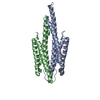


 PDBj
PDBj

 集合体
集合体

 タイプ: L-peptide linking / 分子量: 105.093 Da / 分子数: 2 / 由来タイプ: 合成 / 式: C3H7NO3
 分子量: 18.015 Da / 分子数: 59 / 由来タイプ: 天然 / 式: H2O
 試料調製
試料調製 / ビームライン: BL41XU / 波長: 1 Å
 解析
解析