 Movie
Movie Controller


+ Open data

- Basic information
| Entry | Database: PDB / ID: 3a5x | ||||||
|---|---|---|---|---|---|---|---|
| Title | L-type straight flagellar filament made of full-length flagellin | ||||||
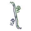
 Components Components | Flagellin | ||||||
 Keywords Keywords | STRUCTURAL PROTEIN / MOTOR PROTEIN / FLAGELLIN / FLAGELLAR FILAMENT / HELICAL RECONSTRUCTION / Bacterial flagellum / Secreted | ||||||
| Function / homology |  Function and homology information Function and homology informationTLR5 cascade / MyD88 cascade initiated on plasma membrane / NFkB and MAPK activation mediated by TRAF6 / The IPAF inflammasome / bacterial-type flagellum / structural molecule activity / extracellular region Similarity search - Function | ||||||
| Biological species |  Salmonella typhimurium (bacteria) Salmonella typhimurium (bacteria) | ||||||
| Method | ELECTRON MICROSCOPY / helical reconstruction / cryo EM / Resolution: 4 Å | ||||||
 Authors Authors | Maki-Yonekura, S. / Yonekura, K. / Namba, K. | ||||||
 Citation Citation | Journal: Nat Struct Mol Biol / Year: 2010 Title: Conformational change of flagellin for polymorphic supercoiling of the flagellar filament. Authors: Saori Maki-Yonekura / Koji Yonekura / Keiichi Namba /  Abstract: The bacterial flagellar filament is a helical propeller rotated by the flagellar motor for bacterial locomotion. The filament is a supercoiled assembly of a single protein, flagellin, and is formed ...The bacterial flagellar filament is a helical propeller rotated by the flagellar motor for bacterial locomotion. The filament is a supercoiled assembly of a single protein, flagellin, and is formed by 11 protofilaments. For bacterial taxis, the reversal of motor rotation switches the supercoil between left- and right-handed, both of which arise from combinations of two distinct conformations and packing interactions of the L-type and R-type protofilaments. Here we report an atomic model of the L-type straight filament by electron cryomicroscopy and helical image analysis. Comparison with the R-type structure shows interesting features: an orientation change of the outer core domains (D1) against the inner core domains (D0) showing almost invariant orientation and packing, a conformational switching within domain D1, and the conformational flexibility of domains D0 and D1 with their spoke-like connection for tight molecular packing. #1: Journal: Nature / Year: 2001Title: Structure of the bacterial flagellar protofilament and implications for a switch for supercoiling. Authors: F A Samatey / K Imada / S Nagashima / F Vonderviszt / T Kumasaka / M Yamamoto / K Namba / Abstract: The bacterial flagellar filament is a helical propeller constructed from 11 protofilaments of a single protein, flagellin. The filament switches between left- and right-handed supercoiled forms when ...The bacterial flagellar filament is a helical propeller constructed from 11 protofilaments of a single protein, flagellin. The filament switches between left- and right-handed supercoiled forms when bacteria switch their swimming mode between running and tumbling. Supercoiling is produced by two different packing interactions of flagellin called L and R. In switching from L to R, the intersubunit distance ( approximately 52 A) along the protofilament decreases by 0.8 A. Changes in the number of L and R protofilaments govern supercoiling of the filament. Here we report the 2.0 A resolution crystal structure of a Salmonella flagellin fragment of relative molecular mass 41,300. The crystal contains pairs of antiparallel straight protofilaments with the R-type repeat. By simulated extension of the protofilament model, we have identified possible switch regions responsible for the bi-stable mechanical switch that generates the 0.8 A difference in repeat distance. #2: Journal: Nature / Year: 2003Title: Complete atomic model of the bacterial flagellar filament by electron cryomicroscopy. Authors: Koji Yonekura / Saori Maki-Yonekura / Keiichi Namba / Abstract: The bacterial flagellar filament is a helical propeller for bacterial locomotion. It is a helical assembly of a single protein, flagellin, and its tubular structure is formed by 11 protofilaments in ...The bacterial flagellar filament is a helical propeller for bacterial locomotion. It is a helical assembly of a single protein, flagellin, and its tubular structure is formed by 11 protofilaments in two distinct conformations, L- and R-type, for supercoiling. The X-ray crystal structure of a flagellin fragment lacking about 100 terminal residues revealed the protofilament structure, but the full filament structure is still essential for understanding the mechanism of supercoiling and polymerization. Here we report a complete atomic model of the R-type filament by electron cryomicroscopy. A density map obtained from image data up to 4 A resolution shows the feature of alpha-helical backbone and some large side chains. The atomic model built on the map reveals intricate molecular packing and an alpha-helical coiled coil formed by the terminal chains in the inner core of the filament, with its intersubunit hydrophobic interactions having an important role in stabilizing the filament. | ||||||
| History |
| ||||||
| Remark 650 | HELIX DETERMINATION METHOD: AUTHOR DETERMINED | ||||||
| Remark 700 | SHEET DETERMINATION METHOD: AUTHOR DETERMINED |
- Structure visualization
| Movie |
Movie viewer |
|---|---|
| Structure viewer | Molecule: MolmilJmol/JSmol |
- Downloads & links
-Download
| PDBx/mmCIF format | 3a5x.cif.gz | 88.9 KB | Display | PDBx/mmCIF format |
|---|---|---|---|---|
| PDB format | pdb3a5x.ent.gz | 65.1 KB | Display | PDB format |
| PDBx/mmJSON format | 3a5x.json.gz | Tree view | PDBx/mmJSON format | |
| Others |  Other downloads Other downloads |
-Validation report
| Summary document | 3a5x_validation.pdf.gz | 546.9 KB | Display | wwPDB validaton report |
|---|---|---|---|---|
| Full document | 3a5x_full_validation.pdf.gz | 560.1 KB | Display | |
| Data in XML | 3a5x_validation.xml.gz | 17.2 KB | Display | |
| Data in CIF | 3a5x_validation.cif.gz | 26.9 KB | Display | |
| Arichive directory | https://data.pdbj.org/pub/pdb/validation_reports/a5/3a5xftp://data.pdbj.org/pub/pdb/validation_reports/a5/3a5x | HTTPS FTP |
-Related structure data
| Related structure data | 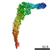 1641MC M: map data used to model this data C: citing same article ( |
|---|---|
| Similar structure data |

-Links
 PDBj
PDBj

- Assembly
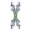
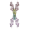
| Deposited unit | 
|
|---|---|
| 1 |
|
-Components
| #1: Protein | Mass: 51537.180 Da / Num. of mol.: 1 / Source method: isolated from a natural source / Source: (natural) Salmonella typhimurium (bacteria) / Strain: SJW1660 / References: UniProt: P06179 |
|---|---|
| Sequence details | A NATURAL MUTATION AT THIS POSITION |
-Experimental details
-Experiment
| Experiment | Method: ELECTRON MICROSCOPY |
|---|---|
| EM experiment | Aggregation state: FILAMENT / 3D reconstruction method: helical reconstruction |
- Sample preparation
| Component | Name: L-TYPE STRAIGHT FLAGELLAR FILAMENT / Type: COMPLEX |
|---|---|
| Buffer solution | Name: 150MM NACL, 2MM MGCL2, 20MM TRIS-HCL, 2-5% GLYCEROL / pH: 7.8 Details: 150MM NACL, 2MM MGCL2, 20MM TRIS-HCL, 2-5% GLYCEROL |
| Specimen | Conc.: 0.1 mg/ml / Embedding applied: NO / Shadowing applied: NO / Staining applied: NO / Vitrification applied: YES |
| Specimen support | Details: QUANTIFOIL R1.2/1.3 (25 NM THICK) |
| Vitrification | Cryogen name: ETHANE |
- Electron microscopy imaging
| Microscopy | Model: JEOL 3000SFF |
|---|---|
| Electron gun | Electron source:  FIELD EMISSION GUN / Accelerating voltage: 300 kV / Illumination mode: FLOOD BEAM FIELD EMISSION GUN / Accelerating voltage: 300 kV / Illumination mode: FLOOD BEAM |
| Electron lens | Mode: BRIGHT FIELD / Nominal magnification: 50000 X / Calibrated magnification: 50000 X / Nominal defocus max: 2220 nm / Nominal defocus min: 1080 nm / Cs: 1.6 mm |
| Specimen holder | Temperature: 4 K / Tilt angle max: 0 ° / Tilt angle min: 0 ° |
| Image recording | Electron dose: 20 e/Å2 / Film or detector model: KODAK SO-163 FILM |
| Radiation | Protocol: SINGLE WAVELENGTH / Monochromatic (M) / Laue (L): M / Scattering type: x-ray |
| Radiation wavelength | Relative weight: 1 |
- Processing
| Software | Name: FEX-PLOR / Classification: refinement | ||||||||||||
|---|---|---|---|---|---|---|---|---|---|---|---|---|---|
| EM software |
| ||||||||||||
| CTF correction | Details: Both amplitude and phase | ||||||||||||
| 3D reconstruction | Method: Helical reconstruction / Resolution: 4 Å / Nominal pixel size: 1 Å / Actual pixel size: 1 Å Magnification calibration: L-TYPE STRAIGHT flagellar filament Details: The layer-line spacing of the L-type straight flagellar filament is more than twice longer than that of the R-type, and frozen-hydrated L-type filaments tended to be more curved. Hence, ...Details: The layer-line spacing of the L-type straight flagellar filament is more than twice longer than that of the R-type, and frozen-hydrated L-type filaments tended to be more curved. Hence, straightening of images using bi-cubic spline was carried out at the first stage. The three-dimensional map was calculated by helical image reconstruction. An initial map produced from 70 filament images, corresponding to 51,272 molecular images, showed relatively poor connections of the ND0 helix and the NS loop. Hence, images showing an inter-particle phase residual against the initial average larger than 40 were excluded from the data set to be averaged. As a result, a three-dimensional map of a better quality was obtained. The number of filament images used for the final image reconstruction was 47, and the total number of molecular images was 36,842, which is consistent with an estimated number of molecular images required to achieve this resolution range. Symmetry type: HELICAL | ||||||||||||
| Atomic model building | Protocol: FLEXIBLE FIT / Space: RECIPROCAL / Target criteria: AMPLITUDE-WEIGHTED PHASE RESIDUAL Details: REFINEMENT PROTOCOL--POSITIONAL AND SIMULATED ANNEALING | ||||||||||||
| Refinement | Highest resolution: 4 Å | ||||||||||||
| Refinement step | Cycle: LAST / Highest resolution: 4 Å
|
